 Movie
Movie Controller

[English] 日本語
 Yorodumi
- PDB-1co6: CRYSTAL STRUCTURE OF FERROCYTOCHROME C2 FROM RHODOPSEUDOMONAS VIRIDIS -
+ Open data

- Basic information
| Entry | Database: PDB / ID: 1co6 | |||||||||
|---|---|---|---|---|---|---|---|---|---|---|
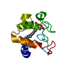
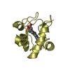
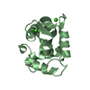
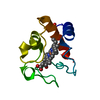
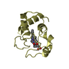
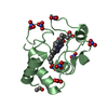
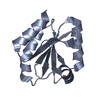
| Title | CRYSTAL STRUCTURE OF FERROCYTOCHROME C2 FROM RHODOPSEUDOMONAS VIRIDIS | |||||||||
 Components Components | PROTEIN (CYTOCHROME C2) | |||||||||
 Keywords Keywords | ELECTRON TRANSPORT / ELECTRON TRANSPORT(HEME PROTEIN) | |||||||||
| Function / homology |  Function and homology information Function and homology informationphotosynthesis / electron transfer activity / heme binding / metal ion binding Similarity search - Function | |||||||||
| Biological species |  Blastochloris viridis (bacteria) Blastochloris viridis (bacteria) | |||||||||
| Method |  X-RAY DIFFRACTION / SYNCHROTRON / MOLECULAR REPLACEMENT / Resolution: 1.6 Å X-RAY DIFFRACTION / SYNCHROTRON / MOLECULAR REPLACEMENT / Resolution: 1.6 Å | |||||||||
 Authors Authors | Miki, K. / Sogabe, S. | |||||||||
 Citation Citation | Journal: J.Mol.Biol. / Year: 1995 Title: Refined crystal structure of ferrocytochrome c2 from Rhodopseudomonas viridis at 1.6 A resolution. Authors: Sogabe, S. / Miki, K. #1: Journal: Acta Crystallogr.,Sect.D / Year: 1994Title: Application of Automatic Molecular-Replacement Procedure to Crystal Structure Analysis of Cytochrrome c2 from Rhodopseudomonas Viridis Authors: Miki, K. / Sogabe, S. / Uno, A. / Ezoe, T. / Kasai, N. / Saeda, M. / Matsuura, Y. / Miki, M. #2: Journal: FEBS Lett. / Year: 1994Title: Structure Similarity of Cytochrrome c2 from Rhodopseudomonas Viridis to Mitochondrial Cytochrome C Revealed by its Crystal Structure at 2.7 A Resokution Authors: Sogabe, S. / Ezoe, T. / Kasai, N. / Saeda, M. / Uno, A. / Miki, M. / Miki, K. | |||||||||
| History |
|
- Structure visualization
| Structure viewer | Molecule: MolmilJmol/JSmol |
|---|
- Downloads & links
-Download
| PDBx/mmCIF format | 1co6.cif.gz | 39.2 KB | Display | PDBx/mmCIF format |
|---|---|---|---|---|
| PDB format | pdb1co6.ent.gz | 26.1 KB | Display | PDB format |
| PDBx/mmJSON format | 1co6.json.gz | Tree view | PDBx/mmJSON format | |
| Others |  Other downloads Other downloads |
-Validation report
| Arichive directory | https://data.pdbj.org/pub/pdb/validation_reports/co/1co6ftp://data.pdbj.org/pub/pdb/validation_reports/co/1co6 | HTTPS FTP |
|---|
-Related structure data
| Related structure data | 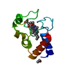 5cytS S: Starting model for refinement |
|---|---|
| Similar structure data |


-Links
 PDBj
PDBj


- Assembly
| Deposited unit | 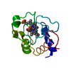
| ||||||||||
|---|---|---|---|---|---|---|---|---|---|---|---|
| 1 |
| ||||||||||
| Unit cell |
|
-Components
| #1: Protein | Mass: 11667.220 Da / Num. of mol.: 1 / Source method: isolated from a natural source / Source: (natural) Blastochloris viridis (bacteria) / References: UniProt: P00083 |
|---|---|
| #2: Chemical | ChemComp-HEC /   Mass: 618.503 Da / Num. of mol.: 1 / Source method: obtained synthetically / Formula: C34H34FeN4O4 Mass: 618.503 Da / Num. of mol.: 1 / Source method: obtained synthetically / Formula: C34H34FeN4O4 |
| #3: Water | ChemComp-HOH /  Mass: 18.015 Da / Num. of mol.: 145 / Source method: isolated from a natural source / Formula: H2O Mass: 18.015 Da / Num. of mol.: 145 / Source method: isolated from a natural source / Formula: H2O |
| Has protein modification | Y |
-Experimental details
-Experiment
| Experiment | Method: X-RAY DIFFRACTION / Number of used crystals: 1 |
|---|
- Sample preparation
| Crystal | Density Matthews: 2.37 Å3/Da / Density % sol: 57.52 % | |||||||||||||||||||||||||
|---|---|---|---|---|---|---|---|---|---|---|---|---|---|---|---|---|---|---|---|---|---|---|---|---|---|---|
| Crystal grow | Method: vapor diffusion, hanging drop / pH: 8.5 Details: CRYSTALS WERE GROWN FROM AN AMMONIUM SULFATE SOLUTION AT PH 8.5, VAPOR DIFFUSION, HANGING DROP | |||||||||||||||||||||||||
| Crystal grow | *PLUS Temperature: 4 ℃ / Method: vapor diffusion, sitting drop | |||||||||||||||||||||||||
| Components of the solutions | *PLUS
|
-Data collection
| Diffraction | Mean temperature: 291 K |
|---|---|
| Diffraction source | Source: SYNCHROTRON / Site: Photon Factory  / Beamline: BL-6A / Wavelength: 1 / Beamline: BL-6A / Wavelength: 1 |
| Detector | Type: WEISSENBERG / Detector: DIFFRACTOMETER / Date: Jul 15, 1994 |
| Radiation | Monochromator: SI(111) / Protocol: SINGLE WAVELENGTH / Monochromatic (M) / Laue (L): M / Scattering type: x-ray |
| Radiation wavelength | Wavelength: 1 Å / Relative weight: 1 |
| Reflection | Resolution: 1.6→20 Å / Num. obs: 16166 / % possible obs: 89.1 % / Observed criterion σ(I): 1 / Redundancy: 1 % / Rmerge(I) obs: 0.072 |
- Processing
| Software |
| ||||||||||||||||||||||||||||||||||||||||||||||||||||||||||||||||||||||||||||||||||||
|---|---|---|---|---|---|---|---|---|---|---|---|---|---|---|---|---|---|---|---|---|---|---|---|---|---|---|---|---|---|---|---|---|---|---|---|---|---|---|---|---|---|---|---|---|---|---|---|---|---|---|---|---|---|---|---|---|---|---|---|---|---|---|---|---|---|---|---|---|---|---|---|---|---|---|---|---|---|---|---|---|---|---|---|---|---|
| Refinement | Method to determine structure: MOLECULAR REPLACEMENT Starting model: 5CYT Resolution: 1.6→6 Å / σ(F): 1 / Details: INITIAL REFINEMENT WITH X-PLOR 3.1
| ||||||||||||||||||||||||||||||||||||||||||||||||||||||||||||||||||||||||||||||||||||
| Refinement step | Cycle: LAST / Resolution: 1.6→6 Å
| ||||||||||||||||||||||||||||||||||||||||||||||||||||||||||||||||||||||||||||||||||||
| Refine LS restraints |
|
