 Movie
Movie Controller

[English] 日本語
 Yorodumi
- PDB-6q6u: Crystal structure of C39S mutant of thioredoxin h1 from Chlamydom... -
+ Open data

- Basic information
| Entry | Database: PDB / ID: 6q6u | ||||||
|---|---|---|---|---|---|---|---|
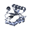
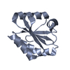
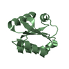
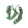
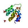
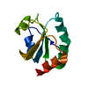
| Title | Crystal structure of C39S mutant of thioredoxin h1 from Chlamydomonas reinhardtii | ||||||
 Components Components | Thioredoxin H-type | ||||||
 Keywords Keywords | ELECTRON TRANSPORT / alpha/beta protein / thioredoxin fold / disulphide oxidoreductase / cell redox homeostatis | ||||||
| Function / homology |  Function and homology information Function and homology information | ||||||
| Biological species |   Chlamydomonas reinhardtii (plant) Chlamydomonas reinhardtii (plant) | ||||||
| Method |  X-RAY DIFFRACTION / SYNCHROTRON / MOLECULAR REPLACEMENT / Resolution: 1.81 Å X-RAY DIFFRACTION / SYNCHROTRON / MOLECULAR REPLACEMENT / Resolution: 1.81 Å | ||||||
 Authors Authors | Fermani, S. / Zaffagnini, M. / Lemaire, S.D. | ||||||
| Funding support |  Italy, 1items Italy, 1items
| ||||||
 Citation Citation | Journal: Antioxidants (Basel) / Year: 2019 Title: Structural and Biochemical Insights into the Reactivity of Thioredoxin h1 fromChlamydomonas reinhardtii. Authors: Marchand, C.H. / Fermani, S. / Rossi, J. / Gurrieri, L. / Tedesco, D. / Henri, J. / Sparla, F. / Trost, P. / Lemaire, S.D. / Zaffagnini, M. | ||||||
| History |
|
- Structure visualization
| Structure viewer | Molecule: MolmilJmol/JSmol |
|---|
- Downloads & links
-Download
| PDBx/mmCIF format | 6q6u.cif.gz | 56.5 KB | Display | PDBx/mmCIF format |
|---|---|---|---|---|
| PDB format | pdb6q6u.ent.gz | 40.5 KB | Display | PDB format |
| PDBx/mmJSON format | 6q6u.json.gz | Tree view | PDBx/mmJSON format | |
| Others |  Other downloads Other downloads |
-Validation report
| Arichive directory | https://data.pdbj.org/pub/pdb/validation_reports/q6/6q6uftp://data.pdbj.org/pub/pdb/validation_reports/q6/6q6u | HTTPS FTP |
|---|
-Related structure data
| Related structure data |  6q46C  6q47C  6q6tC  6q6vC 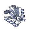 1ep7S S: Starting model for refinement C: citing same article ( |
|---|---|
| Similar structure data |


-Links
 PDBj
PDBj

- Assembly
| Deposited unit | 
| ||||||||
|---|---|---|---|---|---|---|---|---|---|
| 1 | 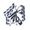
| ||||||||
| 2 | 
| ||||||||
| Unit cell |
| ||||||||
| Components on special symmetry positions |
|
-Components
| #1: Protein | Mass: 11842.665 Da / Num. of mol.: 2 / Mutation: C39S Source method: isolated from a genetically manipulated source Source: (gene. exp.) Chlamydomonas reinhardtii (plant) / Gene: TRXH / Organ: cytoplasm / Production host:  #2: Water | ChemComp-HOH / |  Mass: 18.015 Da / Num. of mol.: 162 / Source method: isolated from a natural source / Formula: H2O Mass: 18.015 Da / Num. of mol.: 162 / Source method: isolated from a natural source / Formula: H2O |
|---|
-Experimental details
-Experiment
| Experiment | Method: X-RAY DIFFRACTION / Number of used crystals: 1 |
|---|
- Sample preparation
| Crystal | Density Matthews: 2.09 Å3/Da / Density % sol: 41.07 % |
|---|---|
| Crystal grow | Temperature: 293 K / Method: vapor diffusion, hanging drop / pH: 6.5 / Details: 10% (w/v) PEG 8K, 10% (w/v) PEG 10K, 0.1 M MES |
-Data collection
| Diffraction | Mean temperature: 100 K / Serial crystal experiment: N |
|---|---|
| Diffraction source | Source: SYNCHROTRON / Site: ELETTRA / Beamline: 5.2R / Wavelength: 1 Å |
| Detector | Type: DECTRIS PILATUS3 S 6M / Detector: PIXEL / Date: May 8, 2014 / Details: Silicon toroidal mirror coated with Rhodium |
| Radiation | Monochromator: Silicon (1 1 1) channel-cut / Protocol: SINGLE WAVELENGTH / Monochromatic (M) / Laue (L): M / Scattering type: x-ray |
| Radiation wavelength | Wavelength: 1 Å / Relative weight: 1 |
| Reflection | Resolution: 1.81→41.94 Å / Num. obs: 18676 / % possible obs: 99.6 % / Observed criterion σ(F): 3 / Observed criterion σ(I): -3 / Redundancy: 5.8 % / Biso Wilson estimate: 24 Å2 / CC1/2: 0.99 / Rmerge(I) obs: 0.111 / Rpim(I) all: 0.057 / Rrim(I) all: 0.133 / Net I/σ(I): 9.7 |
| Reflection shell | Resolution: 1.81→1.84 Å / Redundancy: 5.7 % / Rmerge(I) obs: 0.74 / Mean I/σ(I) obs: 1.9 / Num. unique obs: 1044 / CC1/2: 0.715 / Rpim(I) all: 0.345 / Rrim(I) all: 0.891 / % possible all: 97 |
- Processing
| Software |
| ||||||||||||||||||||||||||||||||||||||||||||||||||||||||||||||||||||||||||||||||||||||||||||||||||
|---|---|---|---|---|---|---|---|---|---|---|---|---|---|---|---|---|---|---|---|---|---|---|---|---|---|---|---|---|---|---|---|---|---|---|---|---|---|---|---|---|---|---|---|---|---|---|---|---|---|---|---|---|---|---|---|---|---|---|---|---|---|---|---|---|---|---|---|---|---|---|---|---|---|---|---|---|---|---|---|---|---|---|---|---|---|---|---|---|---|---|---|---|---|---|---|---|---|---|---|
| Refinement | Method to determine structure: MOLECULAR REPLACEMENT Starting model: 1EP7 Resolution: 1.81→41.94 Å / SU ML: 0.29 / Cross valid method: FREE R-VALUE / σ(F): 0.16 / Phase error: 28.13
| ||||||||||||||||||||||||||||||||||||||||||||||||||||||||||||||||||||||||||||||||||||||||||||||||||
| Solvent computation | Shrinkage radii: 0.9 Å / VDW probe radii: 1.11 Å | ||||||||||||||||||||||||||||||||||||||||||||||||||||||||||||||||||||||||||||||||||||||||||||||||||
| Displacement parameters | Biso mean: 24.7 Å2 | ||||||||||||||||||||||||||||||||||||||||||||||||||||||||||||||||||||||||||||||||||||||||||||||||||
| Refinement step | Cycle: LAST / Resolution: 1.81→41.94 Å
| ||||||||||||||||||||||||||||||||||||||||||||||||||||||||||||||||||||||||||||||||||||||||||||||||||
| Refine LS restraints |
| ||||||||||||||||||||||||||||||||||||||||||||||||||||||||||||||||||||||||||||||||||||||||||||||||||
| LS refinement shell |
|
