 Movie
Movie Controller

[English] 日本語
 Yorodumi
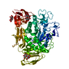
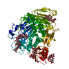
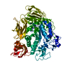
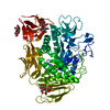
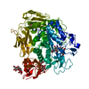
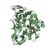
- PDB-1cgt: STRUCTURE OF CYCLODEXTRIN GLYCOSYLTRANSFERASE REFINED AT 2.0 ANGS... -
+ Open data

- Basic information
| Entry | Database: PDB / ID: 1cgt | ||||||
|---|---|---|---|---|---|---|---|
| Title | STRUCTURE OF CYCLODEXTRIN GLYCOSYLTRANSFERASE REFINED AT 2.0 ANGSTROMS RESOLUTION | ||||||
 Components Components | CYCLODEXTRIN GLYCOSYL-TRANSFERASE | ||||||
 Keywords Keywords |  GLYCOSYLTRANSFERASE GLYCOSYLTRANSFERASE | ||||||
| Function / homology |  Function and homology informationcyclomaltodextrin glucanotransferase / cyclomaltodextrin glucanotransferase activity / starch binding / alpha-amylase activity / carbohydrate metabolic process / extracellular region / metal ion binding Function and homology informationcyclomaltodextrin glucanotransferase / cyclomaltodextrin glucanotransferase activity / starch binding / alpha-amylase activity / carbohydrate metabolic process / extracellular region / metal ion bindingSimilarity search - Function | ||||||
| Biological species |  Bacillus circulans (bacteria) Bacillus circulans (bacteria) | ||||||
| Method | X-RAY DIFFRACTION / Resolution: 2 Å | ||||||
 Authors Authors | Klein, C. / Schulz, G.E. | ||||||
 Citation Citation | Journal: J.Mol.Biol. / Year: 1991 Title: Structure of cyclodextrin glycosyltransferase refined at 2.0 A resolution. Authors: Klein, C. / Schulz, G.E. #1: Journal: Protein Eng. / Year: 1990Title: Engineering a Heavy Atom Derivative for the X-Ray Structure Analysis of Cyclodextrin Glycosyltransferase Authors: Klein, C. / Vogel, W. / Bender, H. / Schulz, G.E. #2: Journal: Appl.Microbiol.Biotechnol. / Year: 1990Title: Molecular Cloning, Nucleotide Sequence and Expression in Escherichia Coli of the Beta-Cyclodextrin Glycosyltransferase Gene from Bacillus Circulans Strain No. 8 Authors: Nitschke, L. / Heeger, K. / Bender, H. / Schulz, G.E. #3: Journal: J.Mol.Biol. / Year: 1989Title: Three-Dimensional Structure of Cyclodextrin Glycosyltransferase from Bacillus Circulans at 3.4 Angstroms Resolution Authors: Hofmann, B.E. / Bender, H. / Schulz, G.E. | ||||||
| History |
| ||||||
| Remark 650 | HELIX HELIX STRAND H1 IS A NONHELICAL SEGMENT BETWEEN ASP 63 AND ASN 64. | ||||||
| Remark 700 | SHEET THIS MOLECULE CONTAINS ONE BIFURCATED SHEET. IN ORDER TO REPRESENT THIS FEATURE IN THE SHEET ...SHEET THIS MOLECULE CONTAINS ONE BIFURCATED SHEET. IN ORDER TO REPRESENT THIS FEATURE IN THE SHEET RECORDS BELOW, THE TWO SHEETS, S11 AND S12, ARE DEFINED HAVING STRANDS 1, 2, 3, IN COMMON. |
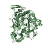
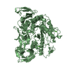
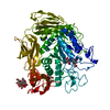
- Structure visualization
| Structure viewer | Molecule: MolmilJmol/JSmol |
|---|
- Downloads & links
-Download
| PDBx/mmCIF format | 1cgt.cif.gz | 152.2 KB | Display | PDBx/mmCIF format |
|---|---|---|---|---|
| PDB format | pdb1cgt.ent.gz | 124 KB | Display | PDB format |
| PDBx/mmJSON format | 1cgt.json.gz | Tree view | PDBx/mmJSON format | |
| Others |  Other downloads Other downloads |
-Validation report
| Arichive directory | https://data.pdbj.org/pub/pdb/validation_reports/cg/1cgtftp://data.pdbj.org/pub/pdb/validation_reports/cg/1cgt | HTTPS FTP |
|---|
-Related structure data
| Similar structure data |
|---|

-Links
 PDBj
PDBj


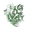
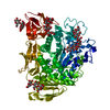
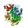
- Assembly
| Deposited unit | 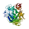
| ||||||||
|---|---|---|---|---|---|---|---|---|---|
| 1 |
| ||||||||
| Unit cell |
| ||||||||
| Atom site foot note | 1: CIS PROLINE - PRO 372 / 2: CIS PROLINE - PRO 505 / 3: CIS PROLINE - PRO 623 / 4: CIS PROLINE - PRO 633 |
-Components
| #1: Protein | Mass: 74565.039 Da / Num. of mol.: 1 Source method: isolated from a genetically manipulated source Source: (gene. exp.) Bacillus circulans (bacteria)References: UniProt: P30920, cyclomaltodextrin glucanotransferase | ||||
|---|---|---|---|---|---|
| #2: Chemical |   Mass: 40.078 Da / Num. of mol.: 2 / Source method: obtained synthetically / Formula: Ca Mass: 40.078 Da / Num. of mol.: 2 / Source method: obtained synthetically / Formula: Ca#3: Water | ChemComp-HOH / | Water Mass: 18.015 Da / Num. of mol.: 588 / Source method: isolated from a natural source / Formula: H2O Mass: 18.015 Da / Num. of mol.: 588 / Source method: isolated from a natural source / Formula: H2OSequence details | THE SEQUENCE BELOW IS THAT DETERMINED | |
-Experimental details
-Experiment
| Experiment | Method: X-RAY DIFFRACTION |
|---|
- Sample preparation
| Crystal | Density Matthews: 3.79 Å3/Da / Density % sol: 67.57 % | |||||||||||||||||||||||||||||||||||||||||||||
|---|---|---|---|---|---|---|---|---|---|---|---|---|---|---|---|---|---|---|---|---|---|---|---|---|---|---|---|---|---|---|---|---|---|---|---|---|---|---|---|---|---|---|---|---|---|---|
| Crystal grow | *PLUS pH: 6.7 / Method: vapor diffusion, hanging drop | |||||||||||||||||||||||||||||||||||||||||||||
| Components of the solutions | *PLUS
|
-Data collection
| Radiation | Scattering type: x-ray |
|---|---|
| Radiation wavelength | Relative weight: 1 |
| Reflection | *PLUS Highest resolution: 2 Å / Lowest resolution: 10 Å / Num. obs: 70862 / % possible obs: 96 % / Num. measured all: 387934 / Rmerge(I) obs: 0.119 |
- Processing
| Software |
| ||||||||||||||||||||||||||||||||||||||||||||||||||||||||||||
|---|---|---|---|---|---|---|---|---|---|---|---|---|---|---|---|---|---|---|---|---|---|---|---|---|---|---|---|---|---|---|---|---|---|---|---|---|---|---|---|---|---|---|---|---|---|---|---|---|---|---|---|---|---|---|---|---|---|---|---|---|---|
| Refinement | Resolution: 2→7 Å /
| ||||||||||||||||||||||||||||||||||||||||||||||||||||||||||||
| Refinement step | Cycle: LAST / Resolution: 2→7 Å
| ||||||||||||||||||||||||||||||||||||||||||||||||||||||||||||
| Refine LS restraints |
| ||||||||||||||||||||||||||||||||||||||||||||||||||||||||||||
| Software | *PLUS Name: X-PLOR / Classification: refinement | ||||||||||||||||||||||||||||||||||||||||||||||||||||||||||||
| Refinement | *PLUS Highest resolution: 2 Å / Lowest resolution: 7 Å / Num. reflection all: 70862 / σ(I): 0 / Rfactor all: 0.166 | ||||||||||||||||||||||||||||||||||||||||||||||||||||||||||||
| Solvent computation | *PLUS | ||||||||||||||||||||||||||||||||||||||||||||||||||||||||||||
| Displacement parameters | *PLUS | ||||||||||||||||||||||||||||||||||||||||||||||||||||||||||||
| Refine LS restraints | *PLUS Type: x_angle_d / Dev ideal: 2.78 |
