- PDB-1bxi: CRYSTAL STRUCTURE OF THE ESCHERICHIA COLI COLICIN E9 DNASE DOMAIN... -
+
Open data
ID or keywords:
Loading...
-
Basic information
Entry
Database: PDB / ID: 1bxi
Title
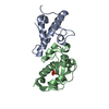
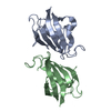
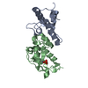
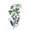
CRYSTAL STRUCTURE OF THE ESCHERICHIA COLI COLICIN E9 DNASE DOMAIN WITH ITS COGNATE IMMUNITY PROTEIN IM9
Components
PROTEIN (COLICIN E9 IMMUNITY PROTEIN)
PROTEIN (COLICIN E9)
Keywords
IMMUNE SYSTEM / COLICINS / ENDONUCLEASE / PROTEIN-PROTEIN INTERACTION
Function / homology
Function and homology information
extrachromosomal circular DNA / bacteriocin immunity / toxic substance binding / endonuclease activity / killing of cells of another organism / Hydrolases; Acting on ester bonds / defense response to bacterium / protein domain specific binding / protein-containing complex / metal ion binding Similarity search - Function
Colicin E7 immunity protein; Chain B, fragment: Endonuclease domain / Colicin/pyocin, DNase domain / Colicin E immunity protein / Colicin immunity protein/pyocin immunity protein / Colicin E immunity protein superfamily / Colicin immunity protein / pyocin immunity protein / Colicin/Pyocin-S2, DNase domain / Colicin/pyocin, DNase domain superfamily / Colicin, receptor domain / Coiled-coil receptor-binding R-domain of colicin E2 ...Colicin E7 immunity protein; Chain B, fragment: Endonuclease domain / Colicin/pyocin, DNase domain / Colicin E immunity protein / Colicin immunity protein/pyocin immunity protein / Colicin E immunity protein superfamily / Colicin immunity protein / pyocin immunity protein / Colicin/Pyocin-S2, DNase domain / Colicin/pyocin, DNase domain superfamily / Colicin, receptor domain / Coiled-coil receptor-binding R-domain of colicin E2 / Cloacin colicin family / Colicin-like bacteriocin tRNase domain / Pyosin/cloacin translocation domain / Pyosin/cloacin translocation domain superfamily / His-Me finger superfamily / Non-ribosomal Peptide Synthetase Peptidyl Carrier Protein; Chain A / Alpha-Beta Complex / Orthogonal Bundle / Mainly Alpha / Alpha Beta Similarity search - Domain/homology
Journal: Thesis / Year: 1998 Title: Crystal Structure of the E.Coli Colicin E9 DNase Domain With its Cognate Immunity Protein Im9 Authors: Kuhlmann, U.C.
In the structure databanks used in Yorodumi, some data are registered as the other names, "COVID-19 virus" and "2019-nCoV". Here are the details of the virus and the list of structure data.
Jan 31, 2019. EMDB accession codes are about to change! (news from PDBe EMDB page)
EMDB accession codes are about to change! (news from PDBe EMDB page)
The allocation of 4 digits for EMDB accession codes will soon come to an end. Whilst these codes will remain in use, new EMDB accession codes will include an additional digit and will expand incrementally as the available range of codes is exhausted. The current 4-digit format prefixed with “EMD-” (i.e. EMD-XXXX) will advance to a 5-digit format (i.e. EMD-XXXXX), and so on. It is currently estimated that the 4-digit codes will be depleted around Spring 2019, at which point the 5-digit format will come into force.
The EM Navigator/Yorodumi systems omit the EMD- prefix.
Related info.:Q: What is EMD? / ID/Accession-code notation in Yorodumi/EM Navigator
Yorodumi is a browser for structure data from EMDB, PDB, SASBDB, etc.
This page is also the successor to EM Navigator detail page, and also detail information page/front-end page for Omokage search.
The word "yorodu" (or yorozu) is an old Japanese word meaning "ten thousand". "mi" (miru) is to see.
Related info.:EMDB / PDB / SASBDB / Comparison of 3 databanks / Yorodumi Search / Aug 31, 2016. New EM Navigator & Yorodumi / Yorodumi Papers / Jmol/JSmol / Function and homology information / Changes in new EM Navigator and Yorodumi
 Movie
Movie Controller

 Yorodumi
 Open data
 Basic information
 Components
 Keywords
 Function and homology information
 X-RAY DIFFRACTION /
  Authors
 Citation
 Structure visualization
 Downloads & links
 Other downloads
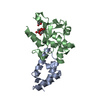

 PDBj
PDBj Assembly
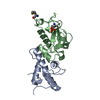

 Mass: 58.693 Da / Num. of mol.: 1 / Source method: obtained synthetically / Formula: Ni
 Mass: 94.971 Da / Num. of mol.: 1 / Source method: obtained synthetically / Formula: PO4
 Mass: 18.015 Da / Num. of mol.: 103 / Source method: isolated from a natural source / Formula: H2O
 Sample preparation
 / Beamline: X31 / Wavelength: 0.979,0.9795,0.9
 Processing