 Movie
Movie Controller

[English] 日本語
 Yorodumi
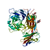
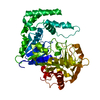
- PDB-1bvn: PIG PANCREATIC ALPHA-AMYLASE IN COMPLEX WITH THE PROTEINACEOUS IN... -
+ Open data

- Basic information
| Entry | Database: PDB / ID: 1bvn | ||||||
|---|---|---|---|---|---|---|---|
| Title | PIG PANCREATIC ALPHA-AMYLASE IN COMPLEX WITH THE PROTEINACEOUS INHIBITOR TENDAMISTAT | ||||||
 Components Components |
| ||||||
 Keywords Keywords | HYDROLASE/HYDROLASE INHIBITOR / GLYCOSYLTRANSFERASE / ALPHA-1 / 4-GLUCAN-4-GLUCANOHYDROLASE / ALPHA-AMYLASE / PROTEINACEOUS ALPHA-AMYLASE INHIBITOR / HYDROLASE-HYDROLASE INHIBITOR COMPLEX | ||||||
| Function / homology |  Function and homology information Function and homology informationalpha-amylase inhibitor activity / alpha-amylase / alpha-amylase activity / carbohydrate catabolic process / chloride ion binding / carbohydrate metabolic process / calcium ion binding / extracellular space Similarity search - Function | ||||||
| Biological species |   Streptomyces tendae (bacteria) Streptomyces tendae (bacteria) | ||||||
| Method |  X-RAY DIFFRACTION / MIR / Resolution: 2.5 Å X-RAY DIFFRACTION / MIR / Resolution: 2.5 Å | ||||||
 Authors Authors | Machius, M. / Wiegand, G. / Epp, O. / Huber, R. | ||||||
 Citation Citation | Journal: J.Mol.Biol. / Year: 1995 Title: The crystal structure of porcine pancreatic alpha-amylase in complex with the microbial inhibitor Tendamistat. Authors: Wiegand, G. / Epp, O. / Huber, R. #1: Journal: J.Mol.Biol. / Year: 1996Title: Carbohydrate and Protein-Based Inhibitors of Porcine Pancreatic Alpha-Amylase: Structure Analysis and Comparison of Their Binding Characteristics Authors: Machius, M. / Vertesy, L. / Huber, R. / Wiegand, G. #2: Journal: Protein Sci. / Year: 1995Title: Carbohydrate Binding Sites in a Pancreatic Alpha-Amylase-Substrate Complex, Derived from X-Ray Structure Analysis at 2.1 Angstrom Resolution Authors: Qian, M. / Haser, R. / Payan, F. #3: Journal: J.Mol.Biol. / Year: 1994Title: Refined Molecular Structure of Pig Pancreatic Alpha-Amylase at 2.1 A Resolution Authors: Larson, S.B. / Greenwood, A. / Cascio, D. / Day, J. / McPherson, A. #4: Journal: Biochemistry / Year: 1994Title: The Active Center of a Mammalian Alpha-Amylase. Structure of the Complex of a Pancreatic Alpha-Amylase with a Carbohydrate Inhibitor Refined to 2.2-A Resolution Authors: Qian, M. / Haser, R. / Buisson, G. / Duee, E. / Payan, F. #5: Journal: J.Mol.Biol. / Year: 1993Title: Structure and Molecular Model Refinement of Pig Pancreatic Alpha-Amylase at 2.1 A Resolution Authors: Qian, M. / Haser, R. / Payan, F. | ||||||
| History |
|
- Structure visualization
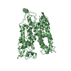
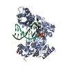
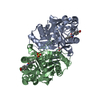
| Structure viewer | Molecule: MolmilJmol/JSmol |
|---|
- Downloads & links
-Download
| PDBx/mmCIF format | 1bvn.cif.gz | 125.9 KB | Display | PDBx/mmCIF format |
|---|---|---|---|---|
| PDB format | pdb1bvn.ent.gz | 97.3 KB | Display | PDB format |
| PDBx/mmJSON format | 1bvn.json.gz | Tree view | PDBx/mmJSON format | |
| Others |  Other downloads Other downloads |
-Validation report
| Arichive directory | https://data.pdbj.org/pub/pdb/validation_reports/bv/1bvnftp://data.pdbj.org/pub/pdb/validation_reports/bv/1bvn | HTTPS FTP |
|---|
-Related structure data
| Similar structure data |
|---|


-Links
 PDBj
PDBj

- Assembly
| Deposited unit | 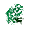
| ||||||||
|---|---|---|---|---|---|---|---|---|---|
| 1 |
| ||||||||
| Unit cell |
|
-Components
| #1: Protein | Mass: 55418.723 Da / Num. of mol.: 1 / Source method: isolated from a natural source / Source: (natural) |
|---|---|
| #2: Protein | Mass: 7967.740 Da / Num. of mol.: 1 / Source method: isolated from a natural source / Source: (natural) Streptomyces tendae (bacteria) / Strain: 4158 / References: UniProt: P01092 |
| #3: Chemical | ChemComp-CA /   Mass: 40.078 Da / Num. of mol.: 1 / Source method: obtained synthetically / Formula: Ca Mass: 40.078 Da / Num. of mol.: 1 / Source method: obtained synthetically / Formula: Ca |
| #4: Chemical | ChemComp-CL /   Mass: 35.453 Da / Num. of mol.: 1 / Source method: obtained synthetically / Formula: Cl Mass: 35.453 Da / Num. of mol.: 1 / Source method: obtained synthetically / Formula: Cl |
| #5: Water | ChemComp-HOH /  Mass: 18.015 Da / Num. of mol.: 161 / Source method: isolated from a natural source / Formula: H2O Mass: 18.015 Da / Num. of mol.: 161 / Source method: isolated from a natural source / Formula: H2O |
| Has protein modification | Y |
| Sequence details | THE SEQUENCE DISCREPANCIES ARE DESCRIBED IN THE CITED REFERENCES GLN1: THIS RESIDUE IS REPORTED TO ...THE SEQUENCE DISCREPANC |
-Experimental details
-Experiment
| Experiment | Method: X-RAY DIFFRACTION / Number of used crystals: 2 |
|---|
- Sample preparation
| Crystal | Density Matthews: 2.5 Å3/Da / Density % sol: 50.5 % | |||||||||||||||||||||||||
|---|---|---|---|---|---|---|---|---|---|---|---|---|---|---|---|---|---|---|---|---|---|---|---|---|---|---|
| Crystal grow | pH: 8 Details: PROTEIN: 12 MG/ML IN 50 MM TRIS/HCL, PH 8.0, MIXED 5:1 WITH 40% (W/V) PEG 1000; RESERVOIR: 0.18-0.20 M SODIUM PHOSPHATE, PH 8.0 HARVESTED IN: 3 M SODIUM ACETATE, PH 7.5 | |||||||||||||||||||||||||
| Crystal | *PLUS | |||||||||||||||||||||||||
| Crystal grow | *PLUS Method: vapor diffusion, sitting drop | |||||||||||||||||||||||||
| Components of the solutions | *PLUS
|
-Data collection
| Diffraction | Mean temperature: 275 K |
|---|---|
| Diffraction source | Source: ROTATING ANODE / Type: RIGAKU RU200 / Wavelength: 1.5418 |
| Detector | Type: KODAK / Detector: FILM / Details: NI-FILTER, DOUBLE-FOCUSING MIRROR SYSTEM |
| Radiation | Monochromator: GRAPHITE / Protocol: SINGLE WAVELENGTH / Monochromatic (M) / Laue (L): M / Scattering type: x-ray |
| Radiation wavelength | Wavelength: 1.5418 Å / Relative weight: 1 |
| Reflection | Resolution: 2.5→29.39 Å / Num. obs: 19789 / % possible obs: 81.3 % / Observed criterion σ(I): 0 / Redundancy: 2.9 % / Biso Wilson estimate: 29.9 Å2 / Rmerge(I) obs: 0.0602 |
| Reflection shell | Resolution: 2.5→2.61 Å / % possible all: 42.4 |
| Reflection | *PLUS Num. measured all: 54641 |
| Reflection shell | *PLUS Rmerge(I) obs: 0.352 |
- Processing
| Software |
| ||||||||||||||||||||||||||||||||||||||||||||||||||||||||||||
|---|---|---|---|---|---|---|---|---|---|---|---|---|---|---|---|---|---|---|---|---|---|---|---|---|---|---|---|---|---|---|---|---|---|---|---|---|---|---|---|---|---|---|---|---|---|---|---|---|---|---|---|---|---|---|---|---|---|---|---|---|---|
| Refinement | Method to determine structure: MIR / Resolution: 2.5→29.4 Å / Data cutoff high absF: 1000000 / Data cutoff low absF: 0.001 / Isotropic thermal model: RESTRAINED / Cross valid method: A POSTERIORI / σ(F): 2 / Details: FINAL RMS COORD. SHIFT 0.0 ANGSTROMS
| ||||||||||||||||||||||||||||||||||||||||||||||||||||||||||||
| Displacement parameters | Biso mean: 26.6 Å2 | ||||||||||||||||||||||||||||||||||||||||||||||||||||||||||||
| Refine analyze | Luzzati coordinate error obs: 0.25 Å | ||||||||||||||||||||||||||||||||||||||||||||||||||||||||||||
| Refinement step | Cycle: LAST / Resolution: 2.5→29.4 Å
| ||||||||||||||||||||||||||||||||||||||||||||||||||||||||||||
| Refine LS restraints |
| ||||||||||||||||||||||||||||||||||||||||||||||||||||||||||||
| LS refinement shell | Resolution: 2.5→2.61 Å / Total num. of bins used: 8
| ||||||||||||||||||||||||||||||||||||||||||||||||||||||||||||
| Xplor file |
| ||||||||||||||||||||||||||||||||||||||||||||||||||||||||||||
| Software | *PLUS Name: X-PLOR / Version: 3.185 / Classification: refinement | ||||||||||||||||||||||||||||||||||||||||||||||||||||||||||||
| Refinement | *PLUS Highest resolution: 2.5 Å / Lowest resolution: 29.4 Å / σ(F): 2 / % reflection Rfree: 5 % / Rfactor Rfree: 0.26 | ||||||||||||||||||||||||||||||||||||||||||||||||||||||||||||
| Solvent computation | *PLUS | ||||||||||||||||||||||||||||||||||||||||||||||||||||||||||||
| Displacement parameters | *PLUS Biso mean: 26.6 Å2 | ||||||||||||||||||||||||||||||||||||||||||||||||||||||||||||
| Refine LS restraints | *PLUS
| ||||||||||||||||||||||||||||||||||||||||||||||||||||||||||||
| LS refinement shell | *PLUS Highest resolution: 2.5 Å / Rfactor Rfree: 0.366 / % reflection Rfree: 5 % / Rfactor Rwork: 0.25 |
