 Movie
Movie Controller


+ Open data

- Basic information
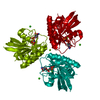
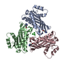
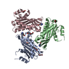
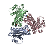
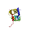
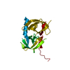
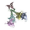
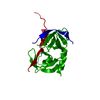
| Entry | Database: PDB / ID: 1ble | ||||||
|---|---|---|---|---|---|---|---|
| Title | PHOSPHOENOLPYRUVATE-DEPENDENT PHOSPHOTRANSFERASE SYSTEM | ||||||
 Components Components | FRUCTOSE PERMEASE | ||||||
 Keywords Keywords | PHOSPHOTRANSFERASE / SUGAR TRANSPORT | ||||||
| Function / homology |  Function and homology information Function and homology informationprotein-Npi-phosphohistidine-D-fructose phosphotransferase / protein-N(PI)-phosphohistidine-sugar phosphotransferase activity / phosphoenolpyruvate-dependent sugar phosphotransferase system / kinase activity / cytoplasm Similarity search - Function | ||||||
| Biological species |  | ||||||
| Method |  X-RAY DIFFRACTION / SYNCHROTRON / MIRAS / Resolution: 2.9 Å X-RAY DIFFRACTION / SYNCHROTRON / MIRAS / Resolution: 2.9 Å | ||||||
 Authors Authors | Schauder, S. / Schirmer, T. | ||||||
 Citation Citation | Journal: J.Mol.Biol. / Year: 1998 Title: Crystal structure of the IIB subunit of a fructose permease (IIBLev) from Bacillus subtilis. Authors: Schauder, S. / Nunn, R.S. / Lanz, R. / Erni, B. / Schirmer, T. #1: Journal: J.Mol.Biol. / Year: 1990Title: Levanase Operon of Bacillus Subtilis Includes a Fructose-Specific Phosphotransferase System Regulating the Expression of the Operon Authors: Martin-Verstraete, I. / Debarbouille, M. / Klier, A. / Rapoport, G. | ||||||
| History |
|
- Structure visualization
| Structure viewer | Molecule: MolmilJmol/JSmol |
|---|
- Downloads & links
-Download
| PDBx/mmCIF format | 1ble.cif.gz | 42.8 KB | Display | PDBx/mmCIF format |
|---|---|---|---|---|
| PDB format | pdb1ble.ent.gz | 30.7 KB | Display | PDB format |
| PDBx/mmJSON format | 1ble.json.gz | Tree view | PDBx/mmJSON format | |
| Others |  Other downloads Other downloads |
-Validation report
| Summary document | 1ble_validation.pdf.gz | 432.4 KB | Display | wwPDB validaton report |
|---|---|---|---|---|
| Full document | 1ble_full_validation.pdf.gz | 434.9 KB | Display | |
| Data in XML | 1ble_validation.xml.gz | 8.3 KB | Display | |
| Data in CIF | 1ble_validation.cif.gz | 10.2 KB | Display | |
| Arichive directory | https://data.pdbj.org/pub/pdb/validation_reports/bl/1bleftp://data.pdbj.org/pub/pdb/validation_reports/bl/1ble | HTTPS FTP |
-Related structure data
| Similar structure data |
|---|
-Links
 PDBj
PDBj- Assembly
| Deposited unit | 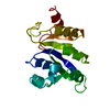
| ||||||||
|---|---|---|---|---|---|---|---|---|---|
| 1 | 
| ||||||||
| Unit cell |
|
-Components
| #1: Protein | Mass: 18241.139 Da / Num. of mol.: 1 Source method: isolated from a genetically manipulated source Source: (gene. exp.) References: UniProt: P26380, protein-Npi-phosphohistidine-sugar phosphotransferase |
|---|---|
| #2: Water | ChemComp-HOH /  Mass: 18.015 Da / Num. of mol.: 7 / Source method: isolated from a natural source / Formula: H2O Mass: 18.015 Da / Num. of mol.: 7 / Source method: isolated from a natural source / Formula: H2O |
-Experimental details
-Experiment
| Experiment | Method: X-RAY DIFFRACTION / Number of used crystals: 1 |
|---|
- Sample preparation
| Crystal | Density Matthews: 2.79 Å3/Da / Density % sol: 56 % | |||||||||||||||||||||||||
|---|---|---|---|---|---|---|---|---|---|---|---|---|---|---|---|---|---|---|---|---|---|---|---|---|---|---|
| Crystal grow | pH: 9 / Details: pH 9.0 | |||||||||||||||||||||||||
| Crystal grow | *PLUS Temperature: 20 ℃ / pH: 7 / Method: vapor diffusion, hanging dropDetails: drop solution was mixed with an equal volume of reservoir solution | |||||||||||||||||||||||||
| Components of the solutions | *PLUS
|
-Data collection
| Diffraction | Mean temperature: 293 K |
|---|---|
| Diffraction source | Source: SYNCHROTRON / Site: SRS  / Beamline: PX9.6 / Wavelength: 0.87 / Beamline: PX9.6 / Wavelength: 0.87 |
| Detector | Type: MARRESEARCH / Detector: IMAGE PLATE / Date: Dec 2, 1995 |
| Radiation | Monochromatic (M) / Laue (L): M / Scattering type: x-ray |
| Radiation wavelength | Wavelength: 0.87 Å / Relative weight: 1 |
| Reflection | Resolution: 2.9→20 Å / Num. obs: 4889 / % possible obs: 97.7 % / Observed criterion σ(I): 3 / Redundancy: 3.5 % / Biso Wilson estimate: 61.6 Å2 / Rmerge(I) obs: 0.054 |
| Reflection shell | Resolution: 2.9→3 Å / Redundancy: 3.6 % / Rmerge(I) obs: 0.288 / % possible all: 96.7 |
| Reflection shell | *PLUS % possible obs: 96.7 % / Mean I/σ(I) obs: 2.5 |
- Processing
| Software |
| ||||||||||||||||||||||||||||||||||||||||||||||||||||||||||||
|---|---|---|---|---|---|---|---|---|---|---|---|---|---|---|---|---|---|---|---|---|---|---|---|---|---|---|---|---|---|---|---|---|---|---|---|---|---|---|---|---|---|---|---|---|---|---|---|---|---|---|---|---|---|---|---|---|---|---|---|---|---|
| Refinement | Method to determine structure: MIRAS / Resolution: 2.9→20 Å / Cross valid method: THROUGHOUT / σ(F): 0 Details: EXCEPT FOR THE ACTIVE LOOP RESIDUE PHE 13, ALL MAIN CHAIN ATOMS ARE WITHIN THE ALLOWED REGIONS OF THE RAMACHANDRAN PLOT.
| ||||||||||||||||||||||||||||||||||||||||||||||||||||||||||||
| Displacement parameters | Biso mean: 64.5 Å2 | ||||||||||||||||||||||||||||||||||||||||||||||||||||||||||||
| Refinement step | Cycle: LAST / Resolution: 2.9→20 Å
| ||||||||||||||||||||||||||||||||||||||||||||||||||||||||||||
| Refine LS restraints |
| ||||||||||||||||||||||||||||||||||||||||||||||||||||||||||||
| LS refinement shell | Resolution: 2.9→3.03 Å / Total num. of bins used: 8
| ||||||||||||||||||||||||||||||||||||||||||||||||||||||||||||
| Software | *PLUS Name: X-PLOR / Version: 3.1 / Classification: refinement | ||||||||||||||||||||||||||||||||||||||||||||||||||||||||||||
| Refine LS restraints | *PLUS
|
