Type: RIGAKU RAXIS IV / Detector: IMAGE PLATE / Date: May 27, 2007 / Details: Varimax-HR
Radiation
Monochromator: Varimax-HR / Protocol: SINGLE WAVELENGTH / Monochromatic (M) / Laue (L): M / Scattering type: x-ray
Radiation wavelength
Wavelength: 1.5418 Å / Relative weight: 1
Reflection
Redundancy: 10.2 % / Av σ(I) over netI: 15.6 / Number: 60706 / Rmerge(I) obs: 0.058 / Χ2: 1.53 / D res high: 2.7 Å / D res low: 50 Å / Num. obs: 5938 / % possible obs: 98.2
Diffraction reflection shell
Highest resolution (Å)
Lowest resolution (Å)
% possible obs (%)
ID
Rmerge(I) obs
Chi squared
Redundancy
5.81
50
99.5
1
0.039
1.919
11.3
4.62
5.81
100
1
0.051
2.106
11.3
4.03
4.62
100
1
0.057
2.408
11.2
3.66
4.03
100
1
0.081
2.295
11
3.4
3.66
100
1
0.099
1.585
11.1
3.2
3.4
100
1
0.104
1.164
11.2
3.04
3.2
100
1
0.124
0.971
11
2.91
3.04
99.8
1
0.144
0.756
10.3
2.8
2.91
95.4
1
0.16
0.508
7.5
2.7
2.8
87.5
1
0.201
0.399
5.6
Reflection
Resolution: 2.7→50 Å / Num. obs: 5938 / % possible obs: 98.2 %
Reflection shell
Resolution: 2.7→2.8 Å / Rmerge(I) obs: 0.201 / % possible all: 87.5
-
Phasing
Phasing MR
Highest resolution
Lowest resolution
Rotation
2.7 Å
29.46 Å
Translation
2.7 Å
29.46 Å
-
Processing
Software
Name
Version
Classification
NB
PHENIX
refinement
SCALEPACK
datascaling
PHASER
phasing
CNS
1.1
refinement
PDB_EXTRACT
2
dataextraction
HKL-2000
datacollection
HKL-2000
datareduction
DENZO
datareduction
Refinement
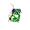
Method to determine structure: MOLECULAR REPLACEMENT Starting model: 2QF0
In the structure databanks used in Yorodumi, some data are registered as the other names, "COVID-19 virus" and "2019-nCoV". Here are the details of the virus and the list of structure data.
Jan 31, 2019. EMDB accession codes are about to change! (news from PDBe EMDB page)
EMDB accession codes are about to change! (news from PDBe EMDB page)
The allocation of 4 digits for EMDB accession codes will soon come to an end. Whilst these codes will remain in use, new EMDB accession codes will include an additional digit and will expand incrementally as the available range of codes is exhausted. The current 4-digit format prefixed with “EMD-” (i.e. EMD-XXXX) will advance to a 5-digit format (i.e. EMD-XXXXX), and so on. It is currently estimated that the 4-digit codes will be depleted around Spring 2019, at which point the 5-digit format will come into force.
The EM Navigator/Yorodumi systems omit the EMD- prefix.
Related info.:Q: What is EMD? / ID/Accession-code notation in Yorodumi/EM Navigator
Yorodumi is a browser for structure data from EMDB, PDB, SASBDB, etc.
This page is also the successor to EM Navigator detail page, and also detail information page/front-end page for Omokage search.
The word "yorodu" (or yorozu) is an old Japanese word meaning "ten thousand". "mi" (miru) is to see.
Related info.:EMDB / PDB / SASBDB / Comparison of 3 databanks / Yorodumi Search / Aug 31, 2016. New EM Navigator & Yorodumi / Yorodumi Papers / Jmol/JSmol / Function and homology information / Changes in new EM Navigator and Yorodumi
 Movie
Movie Controller


 Open data
 Basic information
 Components
 Keywords
 Function and homology information
 X-RAY DIFFRACTION /
  Authors
 Citation
 Structure visualization
 Downloads & links
 Other downloads

 PDBj
PDBj
 Assembly
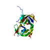
 Sample preparation
 Processing