 Movie
Movie Controller

[English] 日本語
 Yorodumi
- PDB-1bdc: STAPHYLOCOCCUS AUREUS PROTEIN A, IMMUNOGLOBULIN-BINDING B DOMAIN,... -
+ Open data

- Basic information
| Entry | Database: PDB / ID: 1bdc | ||||||
|---|---|---|---|---|---|---|---|
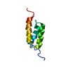
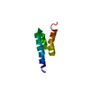
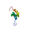
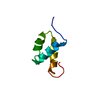
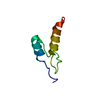
| Title | STAPHYLOCOCCUS AUREUS PROTEIN A, IMMUNOGLOBULIN-BINDING B DOMAIN, NMR, 10 STRUCTURES | ||||||
 Components Components | STAPHYLOCOCCUS AUREUS PROTEIN A | ||||||
 Keywords Keywords | IMMUNOGLOBULIN-BINDING PROTEIN / TRANSMEMBRANE / CELL WALL / IMMUNOGLOBULIN BINDING DOMAIN | ||||||
| Function / homology |  Function and homology information Function and homology information | ||||||
| Biological species |   Staphylococcus aureus (bacteria) Staphylococcus aureus (bacteria) | ||||||
| Method | SOLUTION NMR / HYBRID DISTANCE GEOMETRY-DYNAMICAL SIMULATED ANNEALING METHOD | ||||||
 Authors Authors | Gouda, H. / Torigoe, H. / Saito, A. / Sato, M. / Arata, Y. / Shimada, I. | ||||||
 Citation Citation | Journal: Biochemistry / Year: 1992 Title: Three-dimensional solution structure of the B domain of staphylococcal protein A: comparisons of the solution and crystal structures. Authors: Gouda, H. / Torigoe, H. / Saito, A. / Sato, M. / Arata, Y. / Shimada, I. #1: Journal: FEBS Lett. / Year: 1990Title: 15N Nuclear Magnetic Resonance Studies of the B Domain of Staphylococcal Protein A: Sequence Specific Assignments of the Imide 15N Resonances of the Proline Residues and the Interaction with Human Immunoglobulin G Authors: Torigoe, H. / Shimada, I. / Waelchli, M. / Saito, A. / Sato, M. / Arata, Y. #2: Journal: Biochemistry / Year: 1990Title: Sequential 1H NMR Assignments and Secondary Structure of the B Domain of Staphylococcal Protein A: Structural Changes between the Free B Domain in Solution and the Fc-Bound B Domain in Crystal Authors: Torigoe, H. / Shimada, I. / Saito, A. / Sato, M. / Arata, Y. #3: Journal: Protein Eng. / Year: 1989Title: High Level Expression of a Synthetic Gene Coding for Igg-Binding Domain B of Staphylococcal Protein A Authors: Saito, A. / Honda, S. / Nishi, T. / Koike, M. / Okazaki, K. / Itoh, S. / Sato, M. | ||||||
| History |
|
- Structure visualization
| Structure viewer | Molecule: MolmilJmol/JSmol |
|---|
- Downloads & links
-Download
| PDBx/mmCIF format | 1bdc.cif.gz | 189.8 KB | Display | PDBx/mmCIF format |
|---|---|---|---|---|
| PDB format | pdb1bdc.ent.gz | 154.6 KB | Display | PDB format |
| PDBx/mmJSON format | 1bdc.json.gz | Tree view | PDBx/mmJSON format | |
| Others |  Other downloads Other downloads |
-Validation report
| Summary document | 1bdc_validation.pdf.gz | 351.8 KB | Display | wwPDB validaton report |
|---|---|---|---|---|
| Full document | 1bdc_full_validation.pdf.gz | 440.6 KB | Display | |
| Data in XML | 1bdc_validation.xml.gz | 16.5 KB | Display | |
| Data in CIF | 1bdc_validation.cif.gz | 25 KB | Display | |
| Arichive directory | https://data.pdbj.org/pub/pdb/validation_reports/bd/1bdcftp://data.pdbj.org/pub/pdb/validation_reports/bd/1bdc | HTTPS FTP |
-Related structure data


-Links
 PDBj
PDBj
- Assembly
| Deposited unit | 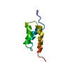
| |||||||||
|---|---|---|---|---|---|---|---|---|---|---|
| 1 |
| |||||||||
| NMR ensembles |
|
-Components
| #1: Protein | Mass: 6778.418 Da / Num. of mol.: 1 / Fragment: B DOMAIN Source method: isolated from a genetically manipulated source Source: (gene. exp.) Staphylococcus aureus (bacteria) / Gene: SYNTHETIC GENE / Plasmid: PPRAFW1 / Production host: |
|---|
-Experimental details
-Experiment
| Experiment | Method: SOLUTION NMR | ||||||||||||||||||||||||||||||||||||
|---|---|---|---|---|---|---|---|---|---|---|---|---|---|---|---|---|---|---|---|---|---|---|---|---|---|---|---|---|---|---|---|---|---|---|---|---|---|
| NMR experiment |
|
 HSQC
HSQC- Sample preparation
| Sample conditions | pH: 5 / Temperature: 303 K |
|---|---|
| Crystal grow | *PLUS Method: other / Details: NMR |
-NMR measurement
| NMR spectrometer | Type: JEOL JNM GSX / Manufacturer: JEOL / Model: JNM GSX / Field strength: 500 MHz |
|---|
- Processing
| Software |
| ||||||||||||
|---|---|---|---|---|---|---|---|---|---|---|---|---|---|
| NMR software |
| ||||||||||||
| Refinement | Method: HYBRID DISTANCE GEOMETRY-DYNAMICAL SIMULATED ANNEALING METHOD Software ordinal: 1 | ||||||||||||
| NMR ensemble | Conformer selection criteria: AT FIRST, THE DEPOSITORS CARRIED OUT THE DISTANCE GEOMETRY CALCULATION BY STARTING FROM 55 INITIAL STRUCTURES. THIS CALCULATION RESULTED IN 41 SOLUTIONS, WHICH HAD ...Conformer selection criteria: AT FIRST, THE DEPOSITORS CARRIED OUT THE DISTANCE GEOMETRY CALCULATION BY STARTING FROM 55 INITIAL STRUCTURES. THIS CALCULATION RESULTED IN 41 SOLUTIONS, WHICH HAD CORRECT POLYPEPTIDE FOLDS EXCLUDING 14 MIRROR-IMAGE SUBSTRUCTURES. NEXT, THE DYNAMICAL SIMULATED ANNEALING CALCULATIONS WERE PERFORMED BY USING THESE 41 SUBSTRUCTURES. THE DISTANCE AND TORSION ANGLE VIOLATIONS OF THE 41 SOLUTIONS OBTAINED BY THE DYNAMICAL SIMULATED ANNEALING CALCULATIONS WERE SMALLER THAN 0.6 ANGSTROMS AND 27 DEGREES, RESPECTIVELY. THE DEPOSITORS SELECTED 10 SOLUTIONS THAT HAD THE DISTANCE AND TORSION ANGLE VIOLATIONS OF SMALLER THAN 0.5 ANGSTROMS AND 10 DEGREES, RESPECTIVELY. Conformers calculated total number: 55 / Conformers submitted total number: 10 |
