 Movie
Movie Controller

[English] 日本語
 Yorodumi
- PDB-1bbl: THREE-DIMENSIONAL SOLUTION STRUCTURE OF THE E3-BINDING DOMAIN OF ... -
+ Open data

- Basic information
| Entry | Database: PDB / ID: 1bbl | ||||||
|---|---|---|---|---|---|---|---|
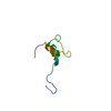
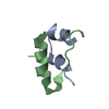
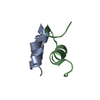
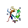
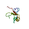
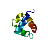
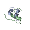
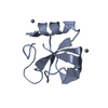
| Title | THREE-DIMENSIONAL SOLUTION STRUCTURE OF THE E3-BINDING DOMAIN OF THE DIHYDROLIPOAMIDE SUCCINYLTRANSFERASE CORE FROM THE 2-OXOGLUTARATE DEHYDROGENASE MULTIENZYME COMPLEX OF ESCHERICHIA COLI | ||||||
 Components Components | DIHYDROLIPOAMIDE SUCCINYLTRANSFERASE | ||||||
 Keywords Keywords | GLYCOLYSIS | ||||||
| Function / homology |  Function and homology information Function and homology informationL-lysine catabolic process to acetyl-CoA via L-saccharopine / dihydrolipoyllysine-residue succinyltransferase / dihydrolipoyllysine-residue succinyltransferase activity / lipoic acid binding / oxoglutarate dehydrogenase complex / tricarboxylic acid cycle / cytoplasm / cytosol Similarity search - Function | ||||||
| Biological species |  | ||||||
| Method | SOLUTION NMR | ||||||
 Authors Authors | Clore, G.M. / Robien, M.A. / Gronenborn, A.M. | ||||||
 Citation Citation | Journal: Biochemistry / Year: 1992 Title: Three-dimensional solution structure of the E3-binding domain of the dihydrolipoamide succinyltransferase core from the 2-oxoglutarate dehydrogenase multienzyme complex of Escherichia coli. Authors: Robien, M.A. / Clore, G.M. / Omichinski, J.G. / Perham, R.N. / Appella, E. / Sakaguchi, K. / Gronenborn, A.M. | ||||||
| History |
|
- Structure visualization
| Structure viewer | Molecule: MolmilJmol/JSmol |
|---|
- Downloads & links
-Download
| PDBx/mmCIF format | 1bbl.cif.gz | 22.2 KB | Display | PDBx/mmCIF format |
|---|---|---|---|---|
| PDB format | pdb1bbl.ent.gz | 14.2 KB | Display | PDB format |
| PDBx/mmJSON format | 1bbl.json.gz | Tree view | PDBx/mmJSON format | |
| Others |  Other downloads Other downloads |
-Validation report
| Arichive directory | https://data.pdbj.org/pub/pdb/validation_reports/bb/1bblftp://data.pdbj.org/pub/pdb/validation_reports/bb/1bbl | HTTPS FTP |
|---|
-Related structure data


-Links
 PDBj
PDBj

- Assembly
| Deposited unit | 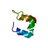
| |||||||||
|---|---|---|---|---|---|---|---|---|---|---|
| 1 |
| |||||||||
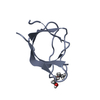
| NMR ensembles |
|
-Components
| #1: Protein | Mass: 5509.133 Da / Num. of mol.: 1 Source method: isolated from a genetically manipulated source Source: (gene. exp.) References: UniProt: P07016, UniProt: P0AFG6*PLUS, dihydrolipoyllysine-residue succinyltransferase |
|---|
-Experimental details
-Experiment
| Experiment | Method: SOLUTION NMR |
|---|
- Sample preparation
| Crystal grow | *PLUS Method: other / Details: NMR |
|---|
- Processing
| Refinement | Software ordinal: 1 Details: DETAILS OF THE STRUCTURE DETERMINATION AND ALL STRUCTURAL STATISTICS ARE GIVEN IN THE PAPER CITED ON *JRNL* RECORDS ABOVE. THE STRUCTURES ARE BASED ON 630 INTERPROTON DISTANCE RESTRAINTS ...Details: DETAILS OF THE STRUCTURE DETERMINATION AND ALL STRUCTURAL STATISTICS ARE GIVEN IN THE PAPER CITED ON *JRNL* RECORDS ABOVE. THE STRUCTURES ARE BASED ON 630 INTERPROTON DISTANCE RESTRAINTS DERIVED FROM NOE MEASUREMENTS; AND 46 PHI AND 35 PSI BACKBONE TORSION ANGLE RESTRAINTS AND 20 CHI1 SIDE CHAIN TORSION ANGLE RESTRAINTS DERIVED FROM COUPLING CONSTANTS AND NOE DATA. THE LATTER ARE OBTAINED USING THE CONFORMATIONAL GRID SEARCH PROGRAM STEREOSEARCH (M.NILGES,G.M.CLORE,A.M.GRONENBORN (1990) BIOPOLYMERS 29, 813-822). THE METHOD USED TO DETERMINE THE STRUCTURES IS THE HYBRID METRIC MATRIX DISTANCE GEOMETRY-DYNAMICAL SIMULATED ANNEALING METHOD (M.NILGES,G.M.CLORE, A.M.GRONENBORN, FEBS LETT. 229, 317-324 (1988)). THIS ENTRY CONTAINS THE SIMULATED ANNEALING RESONANCE MINIMIZED AVERAGE STRUCTURE. THE COORDINATES OF 56 INDIVIDUAL STRUCTURES ARE PRESENTED IN PROTEIN DATA BANK ENTRY 1BAL. (SA)R RESTRAINED MINIMIZED MEAN STRUCTURE WAS DERIVED BY AVERAGING THE COORDINATES OF THE INDIVIDUAL SA STRUCTURES BEST FITTED TO RESIDUES 14 - 30 AND 39 - 47, AND SUBJECTING THE RESULTING COORDINATES TO RESTRAINED MINIMIZATION. RESIDUES 1 - 11 AND 48 - 51 ARE COMPLETELY DISORDERED AND ARE NOT INCLUDED IN THE COORDINATE SET. THE QUANTITY PRESENTED IN COLUMNS 61 - 66 OF THIS ENTRY HAS NO MEANING. THE QUANTITY PRESENTED IN COLUMNS 61 - 66 OF ENTRY 1BAL REPRESENTS THE ATOMIC RMS DEVIATIONS OF THE 56 INDIVIDUAL STRUCTURES ABOUT THE MEAN STRUCTURE. ALL THE INTERPROTON DISTANCE AND TORSION ANGLE RESTRAINTS ARE PRESENTED IN PROTEIN DATA BANK ENTRY R1BBLMR. |
|---|---|
| NMR ensemble | Conformers submitted total number: 1 |
