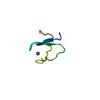
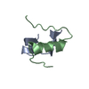
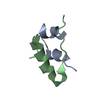
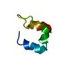
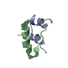
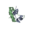
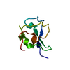
SIGNALING PROTEIN / insulin/relaxin super-family fold
Function / homology
Function and homology information
Relaxin receptors / hormone activity / G protein-coupled receptor binding / G alpha (i) signalling events / G alpha (s) signalling events / extracellular region Similarity search - Function
: / Insulin family / Insulin-like / Insulin/IGF/Relaxin family / Insulin / insulin-like growth factor / relaxin family. / Insulin, conserved site / Insulin family signature. / Insulin-like superfamily Similarity search - Domain/homology
Mass: 3047.586 Da / Num. of mol.: 1 / Fragment: Relaxin 3 B chain / Source method: obtained synthetically Details: This sequence occurs naturally in humans. For this study the peptide has been generated by solid phase peptide synthesis. References: UniProt: Q8WXF3
Mass: 2465.845 Da / Num. of mol.: 1 / Fragment: Relaxin 3 A chain / Source method: obtained synthetically Details: This sequence occurs naturally in humans. For this study the peptide has been generated by solid phase peptide synthesis. References: UniProt: Q8WXF3
Has protein modification
Y
-
Experimental details
-
Experiment
Experiment
Method: SOLUTION NMR
NMR experiment
Conditions-ID
Experiment-ID
Solution-ID
Type
1
1
1
DQF-COSY
1
2
1
2D TOCSY
1
3
1
2D NOESY
1
4
2
2D TOCSY
1
5
2
2D NOESY
NMR details
Text: This structure was determined using standard 2D homonuclear techniques.
-
Sample preparation
Details
Solution-ID
Contents
Solvent system
1
1mM human relaxin-3; 90% H2O, 10% D2O
90% H2O/10% D2O
2
1mM human relaxin-3; 100% D2O
100% D2O
Sample conditions
Conditions-ID
Ionic strength
pH
Pressure (kPa)
Temperature (K)
1
0
3
ambient
298K
2
0
3
ambient
303K
3
0
3
ambient
293K
-
NMR measurement
Radiation
Protocol: SINGLE WAVELENGTH / Monochromatic (M) / Laue (L): M
Radiation wavelength
Relative weight: 1
NMR spectrometer
Type
Manufacturer
Model
Field strength (MHz)
Spectrometer-ID
Bruker AVANCE
Bruker
AVANCE
600
1
Bruker DMX
Bruker
DMX
750
2
-
Processing
NMR software
Name
Version
Developer
Classification
XwinNMR
1.3.5
Bruker
collection
XwinNMR
1.3.5
Bruker
processing
XEASY
1.3.7
Bartels, C. etal.
dataanalysis
CYANA
1
Guntert, P. etal.
structuresolution
CNS
1.1
BrungerA.T. etal.
structuresolution
CNS
1.1
BrungerA.T. etal.
refinement
Refinement
Method: Structures were calculated using torsion angle dynamics, further refined using Cartesian dynamics in explicit solvent. Software ordinal: 1 Details: Structure calculations and refinements were done in CNS using protocols from ARIA.
NMR representative
Selection criteria: lowest energy
NMR ensemble
Conformer selection criteria: structures with the least restraint violations,structures with the lowest energy Conformers calculated total number: 50 / Conformers submitted total number: 20
+
About Yorodumi
-
News
-
Feb 9, 2022. New format data for meta-information of EMDB entries
New format data for meta-information of EMDB entries
Version 3 of the EMDB header file is now the official format.
The previous official version 1.9 will be removed from the archive.
In the structure databanks used in Yorodumi, some data are registered as the other names, "COVID-19 virus" and "2019-nCoV". Here are the details of the virus and the list of structure data.
Jan 31, 2019. EMDB accession codes are about to change! (news from PDBe EMDB page)
EMDB accession codes are about to change! (news from PDBe EMDB page)
The allocation of 4 digits for EMDB accession codes will soon come to an end. Whilst these codes will remain in use, new EMDB accession codes will include an additional digit and will expand incrementally as the available range of codes is exhausted. The current 4-digit format prefixed with “EMD-” (i.e. EMD-XXXX) will advance to a 5-digit format (i.e. EMD-XXXXX), and so on. It is currently estimated that the 4-digit codes will be depleted around Spring 2019, at which point the 5-digit format will come into force.
The EM Navigator/Yorodumi systems omit the EMD- prefix.
Related info.:Q: What is EMD? / ID/Accession-code notation in Yorodumi/EM Navigator
Yorodumi is a browser for structure data from EMDB, PDB, SASBDB, etc.
This page is also the successor to EM Navigator detail page, and also detail information page/front-end page for Omokage search.
The word "yorodu" (or yorozu) is an old Japanese word meaning "ten thousand". "mi" (miru) is to see.
Related info.:EMDB / PDB / SASBDB / Comparison of 3 databanks / Yorodumi Search / Aug 31, 2016. New EM Navigator & Yorodumi / Yorodumi Papers / Jmol/JSmol / Function and homology information / Changes in new EM Navigator and Yorodumi
 Movie
Movie Controller


 Open data
 Basic information
 Components
 Keywords
 Function and homology information
 Authors
 Citation
 Structure visualization
 Downloads & links
 Other downloads
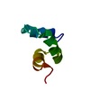
 PDBj
PDBj


 Assembly
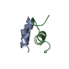
 Sample preparation
 Processing
 CYANA
CYANA