 Movie
Movie Controller

[English] 日本語
 Yorodumi
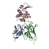
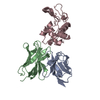
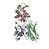
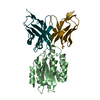
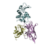
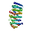
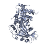
- PDB-1a2y: HEN EGG WHITE LYSOZYME, D18A MUTANT, IN COMPLEX WITH MOUSE MONOCL... -
+ Open data

- Basic information
| Entry | Database: PDB / ID: 1a2y | ||||||
|---|---|---|---|---|---|---|---|
| Title | HEN EGG WHITE LYSOZYME, D18A MUTANT, IN COMPLEX WITH MOUSE MONOCLONAL ANTIBODY D1.3 | ||||||
 Components Components |
| ||||||
 Keywords Keywords | COMPLEX (IMMUNOGLOBULIN/HYDROLASE) / COMPLEX (IMMUNOGLOBULIN-HYDROLASE) / IMMUNOGLOBULIN V REGION /  HYDROLASE / GLYCOSIDASE / BACTERIOLYTIC ENZYME / EGG WHITE / COMPLEX (IMMUNOGLOBULIN-HYDROLASE) complex HYDROLASE / GLYCOSIDASE / BACTERIOLYTIC ENZYME / EGG WHITE / COMPLEX (IMMUNOGLOBULIN-HYDROLASE) complex | ||||||
| Function / homology |  Function and homology informationimmunoglobulin complex / immunoglobulin mediated immune response / antigen binding / Antimicrobial peptides / Neutrophil degranulation / beta-N-acetylglucosaminidase activity / cell wall macromolecule catabolic process / lysozyme / lysozyme activity / killing of cells of another organism ...immunoglobulin complex / immunoglobulin mediated immune response / antigen binding / Antimicrobial peptides / Neutrophil degranulation / beta-N-acetylglucosaminidase activity / cell wall macromolecule catabolic process / lysozyme / lysozyme activity / killing of cells of another organism / defense response to Gram-negative bacterium / adaptive immune response / defense response to Gram-positive bacterium / defense response to bacterium / immune response / endoplasmic reticulum / extracellular space / identical protein binding / cytoplasm Function and homology informationimmunoglobulin complex / immunoglobulin mediated immune response / antigen binding / Antimicrobial peptides / Neutrophil degranulation / beta-N-acetylglucosaminidase activity / cell wall macromolecule catabolic process / lysozyme / lysozyme activity / killing of cells of another organism ...immunoglobulin complex / immunoglobulin mediated immune response / antigen binding / Antimicrobial peptides / Neutrophil degranulation / beta-N-acetylglucosaminidase activity / cell wall macromolecule catabolic process / lysozyme / lysozyme activity / killing of cells of another organism / defense response to Gram-negative bacterium / adaptive immune response / defense response to Gram-positive bacterium / defense response to bacterium / immune response / endoplasmic reticulum / extracellular space / identical protein binding / cytoplasmSimilarity search - Function | ||||||
| Biological species |  Mus musculus (house mouse) Mus musculus (house mouse) Gallus gallus (chicken) Gallus gallus (chicken) | ||||||
| Method | X-RAY DIFFRACTION / SYNCHROTRON / Resolution: 1.5 Å | ||||||
 Authors Authors | Tsuchiya, D. / Mariuzza, R.A. | ||||||
 Citation Citation | Journal: Biochemistry / Year: 1998 Title: A mutational analysis of binding interactions in an antigen-antibody protein-protein complex. Authors: Dall'Acqua, W. / Goldman, E.R. / Lin, W. / Teng, C. / Tsuchiya, D. / Li, H. / Ysern, X. / Braden, B.C. / Li, Y. / Smith-Gill, S.J. / Mariuzza, R.A. #1: Journal: Biochemistry / Year: 1996Title: Hydrogen Bonding and Solvent Structure in an Antigen-Antibody Interface. Crystal Structures and Thermodynamic Characterization of Three Fv Mutants Complexed with Lysozyme Authors: Fields, B.A. / Goldbaum, F.A. / Dall'Acqua, W. / Malchiodi, E.L. / Cauerhff, A. / Schwarz, F.P. / Ysern, X. / Poljak, R.J. / Mariuzza, R.A. #2: Journal: Proc.Natl.Acad.Sci.USA / Year: 1994Title: Bound Water Molecules and Conformational Stabilization Help Mediate an Antigen-Antibody Association Authors: Bhat, T.N. / Bentley, G.A. / Boulot, G. / Greene, M.I. / Tello, D. / Dall'Acqua, W. / Souchon, H. / Schwarz, F.P. / Mariuzza, R.A. / Poljak, R.J. #3: Journal: J.Mol.Biol. / Year: 1994Title: Solvent Rearrangement in an Antigen-Antibody Interface Introduced by Site-Directed Mutagenesis of the Antibody Combining Site Authors: Ysern, X. / Fields, B.A. / Bhat, T.N. / Goldbaum, F.A. / Dall'Acqua, W. / Schwarz, F.P. / Poljak, R.J. / Mariuzza, R.A. | ||||||
| History |
|
- Structure visualization
| Structure viewer | Molecule: MolmilJmol/JSmol |
|---|
- Downloads & links
-Download
| PDBx/mmCIF format | 1a2y.cif.gz | 89.2 KB | Display | PDBx/mmCIF format |
|---|---|---|---|---|
| PDB format | pdb1a2y.ent.gz | 69.9 KB | Display | PDB format |
| PDBx/mmJSON format | 1a2y.json.gz | Tree view | PDBx/mmJSON format | |
| Others |  Other downloads Other downloads |
-Validation report
| Arichive directory | https://data.pdbj.org/pub/pdb/validation_reports/a2/1a2yftp://data.pdbj.org/pub/pdb/validation_reports/a2/1a2y | HTTPS FTP |
|---|
-Related structure data
| Related structure data | 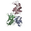 1vfbS S: Starting model for refinement |
|---|---|
| Similar structure data |


-Links
 PDBj
PDBj


- Assembly
| Deposited unit | 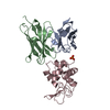
| ||||||||
|---|---|---|---|---|---|---|---|---|---|
| 1 |
| ||||||||
| Unit cell |
|
-Components
| #1: Antibody | Mass: 11700.977 Da / Num. of mol.: 1 Source method: isolated from a genetically manipulated source Source: (gene. exp.) Mus musculus (house mouse) / Organ: EGG / Production host:  Escherichia coli (E. coli) / References: UniProt: P01635 Escherichia coli (E. coli) / References: UniProt: P01635 |
|---|---|
| #2: Antibody | Mass: 12857.275 Da / Num. of mol.: 1 Source method: isolated from a genetically manipulated source Source: (gene. exp.) Mus musculus (house mouse) / Organ: EGG / Production host: Escherichia coli (E. coli) / References: UniProt: P01820 |
| #3: Protein | Mass: 14287.150 Da / Num. of mol.: 1 / Mutation: D18A Source method: isolated from a genetically manipulated source Source: (gene. exp.) Gallus gallus (chicken) / Cell: EGG / Cellular location: CYTOPLASM (WHITE) / Organ: EGG / Production host:  Saccharomyces cerevisiae (brewer's yeast) / References: UniProt: P00698, lysozyme Saccharomyces cerevisiae (brewer's yeast) / References: UniProt: P00698, lysozyme |
| #4: Chemical | ChemComp-PO4 / Phosphate  Mass: 94.971 Da / Num. of mol.: 1 / Source method: obtained synthetically / Formula: PO4 Mass: 94.971 Da / Num. of mol.: 1 / Source method: obtained synthetically / Formula: PO4 |
| #5: Water | ChemComp-HOH / Water Mass: 18.015 Da / Num. of mol.: 485 / Source method: isolated from a natural source / Formula: H2O Mass: 18.015 Da / Num. of mol.: 485 / Source method: isolated from a natural source / Formula: H2O |
-Experimental details
-Experiment
| Experiment | Method: X-RAY DIFFRACTION / Number of used crystals: 1 |
|---|
- Sample preparation
| Crystal | Density Matthews: 2.58 Å3/Da / Density % sol: 52.35 % | |||||||||||||||
|---|---|---|---|---|---|---|---|---|---|---|---|---|---|---|---|---|
| Crystal grow | pH: 6.5 / Details: pH 6.5 | |||||||||||||||
| Crystal grow | *PLUS Method: vapor diffusion, hanging drop | |||||||||||||||
| Components of the solutions | *PLUS
|
-Data collection
| Diffraction | Mean temperature: 100 K |
|---|---|
| Diffraction source | Source: SYNCHROTRON / Site: CHESS  / Type: CHESS / Wavelength: 0.928 / Type: CHESS / Wavelength: 0.928 |
| Detector | Detector: CCD / Date: Jul 1, 1997 |
| Radiation | Monochromatic (M) / Laue (L): M / Scattering type: x-ray |
| Radiation wavelength | Wavelength: 0.928 Å / Relative weight: 1 |
| Reflection | Highest resolution: 1.5 Å / Num. obs: 59595 / % possible obs: 90.4 % / Observed criterion σ(I): 3 / Rmerge(I) obs: 0.069 |
| Reflection shell | Resolution: 1.5→1.55 Å / Rmerge(I) obs: 0.301 / % possible all: 30.1 |
| Reflection | *PLUS Num. measured all: 403840 |
| Reflection shell | *PLUS % possible obs: 50.6 % |
- Processing
| Software |
| ||||||||||||||||||||||||||||||||||||||||||||||||||||||||||||
|---|---|---|---|---|---|---|---|---|---|---|---|---|---|---|---|---|---|---|---|---|---|---|---|---|---|---|---|---|---|---|---|---|---|---|---|---|---|---|---|---|---|---|---|---|---|---|---|---|---|---|---|---|---|---|---|---|---|---|---|---|---|
| Refinement | Starting model: PDB ENTRY 1VFB Resolution: 1.5→7 Å / Data cutoff high absF: 100000 / Data cutoff low absF: 0.1 / Cross valid method: THROUGHOUT / σ(F): 3
| ||||||||||||||||||||||||||||||||||||||||||||||||||||||||||||
| Refine analyze | Luzzati coordinate error obs: 0.2 Å / Luzzati sigma a obs: 0.2 Å | ||||||||||||||||||||||||||||||||||||||||||||||||||||||||||||
| Refinement step | Cycle: LAST / Resolution: 1.5→7 Å
| ||||||||||||||||||||||||||||||||||||||||||||||||||||||||||||
| Refine LS restraints |
| ||||||||||||||||||||||||||||||||||||||||||||||||||||||||||||
| LS refinement shell | Resolution: 1.5→1.55 Å / Total num. of bins used: 10
| ||||||||||||||||||||||||||||||||||||||||||||||||||||||||||||
| Xplor file |
| ||||||||||||||||||||||||||||||||||||||||||||||||||||||||||||
| Software | *PLUS Name: X-PLOR / Version: 3.1 / Classification: refinement | ||||||||||||||||||||||||||||||||||||||||||||||||||||||||||||
| Refine LS restraints | *PLUS
|
