 Movie
Movie Controller


+ Open data

- Basic information
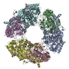
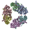
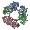
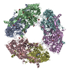
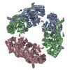
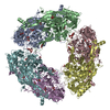
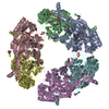
| Entry | Database: PDB / ID: 1a2v | ||||||
|---|---|---|---|---|---|---|---|
| Title | COPPER AMINE OXIDASE FROM HANSENULA POLYMORPHA | ||||||
 Components Components | METHYLAMINE OXIDASE | ||||||
 Keywords Keywords | AMINE OXIDASE / QUINOPROTEIN / TOPAQUINONE ENZYME / TPQ | ||||||
| Function / homology |  Function and homology information Function and homology informationprimary-amine oxidase / primary methylamine oxidase activity / amine metabolic process / quinone binding / peroxisome / copper ion binding Similarity search - Function | ||||||
| Biological species |  Pichia angusta (fungus) Pichia angusta (fungus) | ||||||
| Method |  X-RAY DIFFRACTION / MOLECULAR REPLACEMENT, SIR / Resolution: 2.4 Å X-RAY DIFFRACTION / MOLECULAR REPLACEMENT, SIR / Resolution: 2.4 Å | ||||||
 Authors Authors | Li, R. / Mathews, F.S. | ||||||
 Citation Citation | Journal: Acta Crystallogr.,Sect.D / Year: 1997 Title: Crystallographic study of yeast copper amine oxidase. Authors: Li, R. / Chen, L. / Cai, D. / Klinman, J.P. / Mathews, F.S. | ||||||
| History |
|
- Structure visualization
| Structure viewer | Molecule: MolmilJmol/JSmol |
|---|
- Downloads & links
-Download
| PDBx/mmCIF format | 1a2v.cif.gz | 806.6 KB | Display | PDBx/mmCIF format |
|---|---|---|---|---|
| PDB format | pdb1a2v.ent.gz | 669.2 KB | Display | PDB format |
| PDBx/mmJSON format | 1a2v.json.gz | Tree view | PDBx/mmJSON format | |
| Others |  Other downloads Other downloads |
-Validation report
| Arichive directory | https://data.pdbj.org/pub/pdb/validation_reports/a2/1a2vftp://data.pdbj.org/pub/pdb/validation_reports/a2/1a2v | HTTPS FTP |
|---|
-Related structure data
| Related structure data |  1oacS S: Starting model for refinement |
|---|---|
| Similar structure data |


-Links
 PDBj
PDBj- Assembly
| Deposited unit | 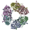
| ||||||||||||||||||||||||
|---|---|---|---|---|---|---|---|---|---|---|---|---|---|---|---|---|---|---|---|---|---|---|---|---|---|
| 1 |
| ||||||||||||||||||||||||
| Unit cell |
| ||||||||||||||||||||||||
| Noncrystallographic symmetry (NCS) | NCS oper:
|
-Components
| #1: Protein | Mass: 73690.047 Da / Num. of mol.: 6 Source method: isolated from a genetically manipulated source Source: (gene. exp.) Pichia angusta (fungus) / Production host:  #2: Chemical | ChemComp-CU /   Mass: 63.546 Da / Num. of mol.: 6 / Source method: obtained synthetically / Formula: Cu Mass: 63.546 Da / Num. of mol.: 6 / Source method: obtained synthetically / Formula: Cu#3: Water | ChemComp-HOH / |  Mass: 18.015 Da / Num. of mol.: 2556 / Source method: isolated from a natural source / Formula: H2O Mass: 18.015 Da / Num. of mol.: 2556 / Source method: isolated from a natural source / Formula: H2O |
|---|
-Experimental details
-Experiment
| Experiment | Method: X-RAY DIFFRACTION / Number of used crystals: 1 |
|---|
- Sample preparation
| Crystal | Density Matthews: 2.6 Å3/Da / Density % sol: 47 % | ||||||||||||||||||||||||||||||
|---|---|---|---|---|---|---|---|---|---|---|---|---|---|---|---|---|---|---|---|---|---|---|---|---|---|---|---|---|---|---|---|
| Crystal grow | pH: 6.2 Details: PROTEIN WAS CRYSTALLIZED IN SITTING DROPS FROM 7-9% PEG 8000 AND 0.3 M POTASSIUM PHOSPHATE BUFFER, PH 6.2 | ||||||||||||||||||||||||||||||
| Crystal grow | *PLUS pH: 6.5 / Method: vapor diffusion, sitting drop | ||||||||||||||||||||||||||||||
| Components of the solutions | *PLUS
|
-Data collection
| Diffraction | Mean temperature: 100 K |
|---|---|
| Diffraction source | Wavelength: 1.5418 |
| Detector | Type: RIGAKU / Detector: IMAGE PLATE / Date: Dec 1, 1995 / Details: MIRRORS |
| Radiation | Monochromator: NI FILTER / Monochromatic (M) / Laue (L): M / Scattering type: x-ray |
| Radiation wavelength | Wavelength: 1.5418 Å / Relative weight: 1 |
| Reflection | Resolution: 2.4→100 Å / Num. obs: 172832 / % possible obs: 87.2 % / Observed criterion σ(I): 0 / Redundancy: 2.8 % / Biso Wilson estimate: 27 Å2 / Rmerge(I) obs: 0.053 / Rsym value: 0.053 / Net I/σ(I): 12.5 |
| Reflection shell | Resolution: 2.4→2.55 Å / Redundancy: 2.8 % / Rmerge(I) obs: 0.247 / Mean I/σ(I) obs: 2.4 / Rsym value: 0.247 / % possible all: 66.8 |
| Reflection shell | *PLUS % possible obs: 66.8 % |
- Processing
| Software |
| ||||||||||||||||||||||||||||||||||||||||||||||||||||||||||||||||||||||||||||||||
|---|---|---|---|---|---|---|---|---|---|---|---|---|---|---|---|---|---|---|---|---|---|---|---|---|---|---|---|---|---|---|---|---|---|---|---|---|---|---|---|---|---|---|---|---|---|---|---|---|---|---|---|---|---|---|---|---|---|---|---|---|---|---|---|---|---|---|---|---|---|---|---|---|---|---|---|---|---|---|---|---|---|
| Refinement | Method to determine structure: MOLECULAR REPLACEMENT, SIR Starting model: PDB ENTRY 1OAC Resolution: 2.4→100 Å / Rfactor Rfree error: 0.003 / Data cutoff high absF: 10000000 / Data cutoff low absF: 0.001 / Isotropic thermal model: RESTRAINED / Cross valid method: THROUGHOUT / σ(F): 1 Details: BULK SOLVENT MODEL USED NCS RESTRAINTS. GROUP 1 POSITIONAL MAIN CHAIN (KCAL/MOL-A**2) : 15 GROUP 1 B-FACTOR MAIN CHAIN (A**2) : 4 GROUP 1 POSITIONAL SIDE CHAIN (KCAL/MOL-A**2) : 10 GROUP 1 B- ...Details: BULK SOLVENT MODEL USED NCS RESTRAINTS. GROUP 1 POSITIONAL MAIN CHAIN (KCAL/MOL-A**2) : 15 GROUP 1 B-FACTOR MAIN CHAIN (A**2) : 4 GROUP 1 POSITIONAL SIDE CHAIN (KCAL/MOL-A**2) : 10 GROUP 1 B-FACTOR MAIN CHAIN (A**2) : 4 GROUP 2 POSITIONAL MAIN CHAIN (KCAL/MOL-A**2) : 15 GROUP 2 B-FACTOR MAIN CHAIN (A**2) : 4 GROUP 2 POSITIONAL SIDE CHAIN (KCAL/MOL-A**2) : 10 GROUP 2 B-FACTOR MAIN CHAIN (A**2) : 4 GROUP 3 POSITIONAL MAIN CHAIN (KCAL/MOL-A**2) : 15 GROUP 3 B-FACTOR MAIN CHAIN (A**2) : 4 GROUP 3 POSITIONAL SIDE CHAIN (KCAL/MOL-A**2) : 10 GROUP 3 B-FACTOR MAIN CHAIN (A**2) : 4 GROUP 4 POSITIONAL MAIN CHAIN (KCAL/MOL-A**2) : 15 GROUP 4 B-FACTOR MAIN CHAIN (A**2) : 4 GROUP 4 POSITIONAL SIDE CHAIN (KCAL/MOL-A**2) : 10 GROUP 4 B-FACTOR MAIN CHAIN (A**2) : 4 GROUP 5 POSITIONAL MAIN CHAIN (KCAL/MOL-A**2) : 15 GROUP 5 B-FACTOR MAIN CHAIN (A**2) : 4 GROUP 5 POSITIONAL SIDE CHAIN (KCAL/MOL-A**2) : 10 GROUP 5 B-FACTOR MAIN CHAIN (A**2) : 4 GROUP 6 POSITIONAL MAIN CHAIN (KCAL/MOL-A**2) : 15 GROUP 6 B-FACTOR MAIN CHAIN (A**2) : 4 GROUP 6 POSITIONAL SIDE CHAIN (KCAL/MOL-A**2) : 10 GROUP 6 B-FACTOR MAIN CHAIN (A**2) : 4
| ||||||||||||||||||||||||||||||||||||||||||||||||||||||||||||||||||||||||||||||||
| Displacement parameters | Biso mean: 24.4 Å2 | ||||||||||||||||||||||||||||||||||||||||||||||||||||||||||||||||||||||||||||||||
| Refine analyze |
| ||||||||||||||||||||||||||||||||||||||||||||||||||||||||||||||||||||||||||||||||
| Refinement step | Cycle: LAST / Resolution: 2.4→100 Å
| ||||||||||||||||||||||||||||||||||||||||||||||||||||||||||||||||||||||||||||||||
| Refine LS restraints |
| ||||||||||||||||||||||||||||||||||||||||||||||||||||||||||||||||||||||||||||||||
| LS refinement shell | Resolution: 2.4→2.55 Å / Rfactor Rfree error: 0.01 / Total num. of bins used: 6
| ||||||||||||||||||||||||||||||||||||||||||||||||||||||||||||||||||||||||||||||||
| Xplor file |
| ||||||||||||||||||||||||||||||||||||||||||||||||||||||||||||||||||||||||||||||||
| Software | *PLUS Name: X-PLOR / Version: 3.843 / Classification: refinement | ||||||||||||||||||||||||||||||||||||||||||||||||||||||||||||||||||||||||||||||||
| Refinement | *PLUS | ||||||||||||||||||||||||||||||||||||||||||||||||||||||||||||||||||||||||||||||||
| Solvent computation | *PLUS | ||||||||||||||||||||||||||||||||||||||||||||||||||||||||||||||||||||||||||||||||
| Displacement parameters | *PLUS | ||||||||||||||||||||||||||||||||||||||||||||||||||||||||||||||||||||||||||||||||
| Refine LS restraints | *PLUS
| ||||||||||||||||||||||||||||||||||||||||||||||||||||||||||||||||||||||||||||||||
| LS refinement shell | *PLUS Rfactor obs: 0.279 |
