 Movie
Movie Controller

[English] 日本語
 Yorodumi
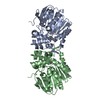
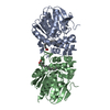
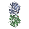
- PDB-5ng7: Novel epoxide hydrolases belonging to the alpha/beta hydrolases s... -
+ Open data

- Basic information
| Entry | Database: PDB / ID: 5ng7 | ||||||
|---|---|---|---|---|---|---|---|
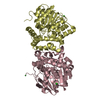
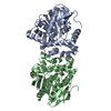
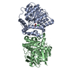
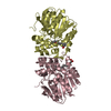
| Title | Novel epoxide hydrolases belonging to the alpha/beta hydrolases superfamily in metagenomes from hot environments | ||||||
 Components Components | epoxide hydrolase | ||||||
 Keywords Keywords | HYDROLASE / Epoxide hydrolases / metagenomics / industrial biocatalysis / stereoselectivity / protein structure | ||||||
| Function / homology | Alpha/Beta hydrolase fold, catalytic domain / Rossmann fold / 3-Layer(aba) Sandwich / Alpha Beta / DI(HYDROXYETHYL)ETHER / SERINE Function and homology information Function and homology information | ||||||
| Biological species | metagenome (others) | ||||||
| Method | X-RAY DIFFRACTION / SYNCHROTRON / MOLECULAR REPLACEMENT / Resolution: 1.39 Å | ||||||
 Authors Authors | Ferrandi, E.E. / De Rose, S.A. / Sayer, C. / Guazzelli, E. / Marchesi, C. / Saneei, V. / Isupov, M.N. / Littlechild, J.A. / Monti, D. | ||||||
| Funding support |  United Kingdom, 1items United Kingdom, 1items
| ||||||
 Citation Citation | Journal: Front Bioeng Biotechnol / Year: 2018 Title: New Thermophilic alpha / beta Class Epoxide Hydrolases Found in Metagenomes From Hot Environments. Authors: Ferrandi, E.E. / Sayer, C. / De Rose, S.A. / Guazzelli, E. / Marchesi, C. / Saneei, V. / Isupov, M.N. / Littlechild, J.A. / Monti, D. | ||||||
| History |
|
- Structure visualization
| Structure viewer | Molecule: MolmilJmol/JSmol |
|---|
- Downloads & links
-Download
| PDBx/mmCIF format | 5ng7.cif.gz | 158.1 KB | Display | PDBx/mmCIF format |
|---|---|---|---|---|
| PDB format | pdb5ng7.ent.gz | 126.3 KB | Display | PDB format |
| PDBx/mmJSON format | 5ng7.json.gz | Tree view | PDBx/mmJSON format | |
| Others |  Other downloads Other downloads |
-Validation report
| Arichive directory | https://data.pdbj.org/pub/pdb/validation_reports/ng/5ng7ftp://data.pdbj.org/pub/pdb/validation_reports/ng/5ng7 | HTTPS FTP |
|---|
-Related structure data
| Related structure data |  5nfqC 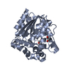 4inzS S: Starting model for refinement C: citing same article ( |
|---|---|
| Similar structure data |


-Links
 PDBj
PDBj- Assembly
| Deposited unit | 
| ||||||||||||||||||
|---|---|---|---|---|---|---|---|---|---|---|---|---|---|---|---|---|---|---|---|
| 1 |
| ||||||||||||||||||
| Unit cell |
| ||||||||||||||||||
| Components on special symmetry positions |
| ||||||||||||||||||
| Noncrystallographic symmetry (NCS) | NCS domain:
NCS domain segments: Component-ID: 0 / Ens-ID: 1 / Beg auth comp-ID: ASN / Beg label comp-ID: ASN / End auth comp-ID: LYS / End label comp-ID: LYS / Refine code: 0 / Auth seq-ID: 2 - 290 / Label seq-ID: 2 - 290
|
-Components
-Protein , 1 types, 2 molecules AB
| #1: Protein | Mass: 34940.449 Da / Num. of mol.: 2 Source method: isolated from a genetically manipulated source Source: (gene. exp.) metagenome (others) / Production host:  Escherichia coli (E. coli) / References: soluble epoxide hydrolase Escherichia coli (E. coli) / References: soluble epoxide hydrolase |
|---|
-Non-polymers , 5 types, 624 molecules 


| #2: Chemical | Chloride Mass: 35.453 Da / Num. of mol.: 3 / Source method: obtained synthetically / Formula: Cl Mass: 35.453 Da / Num. of mol.: 3 / Source method: obtained synthetically / Formula: Cl#3: Chemical | Ethylene glycol Mass: 62.068 Da / Num. of mol.: 3 / Source method: obtained synthetically / Formula: C2H6O2 Mass: 62.068 Da / Num. of mol.: 3 / Source method: obtained synthetically / Formula: C2H6O2#4: Chemical | ChemComp-SER / | Serine Type: L-peptide linking / Mass: 105.093 Da / Num. of mol.: 1 / Source method: obtained synthetically / Formula: C3H7NO3 Type: L-peptide linking / Mass: 105.093 Da / Num. of mol.: 1 / Source method: obtained synthetically / Formula: C3H7NO3#5: Chemical | ChemComp-PEG / | Diethylene glycol Mass: 106.120 Da / Num. of mol.: 1 / Source method: obtained synthetically / Formula: C4H10O3 Mass: 106.120 Da / Num. of mol.: 1 / Source method: obtained synthetically / Formula: C4H10O3#6: Water | ChemComp-HOH / | WaterMass: 18.015 Da / Num. of mol.: 616 / Source method: isolated from a natural source / Formula: H2O |
|---|
-Experimental details
-Experiment
| Experiment | Method: X-RAY DIFFRACTION / Number of used crystals: 1 |
|---|
- Sample preparation
| Crystal | Density Matthews: 1.92 Å3/Da / Density % sol: 35.79 % |
|---|---|
| Crystal grow | Temperature: 291 K / Method: microbatch / pH: 7.5 Details: 200 mM Magnesium formate dehydrate, 100mM Sodium Hepes 7.5 20 % w/v PEG 3350 |
-Data collection
| Diffraction | Mean temperature: 100 K |
|---|---|
| Diffraction source | Source: SYNCHROTRON / Site: Diamond / Beamline: I04 / Wavelength: 0.9795 Å |
| Detector | Type: DECTRIS PILATUS3 6M / Detector: PIXEL / Date: Aug 8, 2016 |
| Radiation | Protocol: SINGLE WAVELENGTH / Monochromatic (M) / Laue (L): M / Scattering type: x-ray |
| Radiation wavelength | Wavelength: 0.9795 Å / Relative weight: 1 |
| Reflection | Resolution: 1.39→39.51 Å / Num. obs: 104819 / % possible obs: 98.2 % / Redundancy: 3.2 % / Biso Wilson estimate: 24.9 Å2 / Rsym value: 0.035 / Net I/σ(I): 15.6 |
| Reflection shell | Resolution: 1.39→1.41 Å / Redundancy: 2.5 % / Mean I/σ(I) obs: 1.2 / Num. unique obs: 4187 / CC1/2: 0.483 / Rsym value: 0.77 / % possible all: 79 |
- Processing
| Software |
| ||||||||||||||||||||||||||||||||||||||||||||||||||||||||||||||||||||||||||||||||||||||||||||||||||||||||||||||||||||||||||||||||||||||||||||||||||||||||||||||||||||||||||||||||||||||
|---|---|---|---|---|---|---|---|---|---|---|---|---|---|---|---|---|---|---|---|---|---|---|---|---|---|---|---|---|---|---|---|---|---|---|---|---|---|---|---|---|---|---|---|---|---|---|---|---|---|---|---|---|---|---|---|---|---|---|---|---|---|---|---|---|---|---|---|---|---|---|---|---|---|---|---|---|---|---|---|---|---|---|---|---|---|---|---|---|---|---|---|---|---|---|---|---|---|---|---|---|---|---|---|---|---|---|---|---|---|---|---|---|---|---|---|---|---|---|---|---|---|---|---|---|---|---|---|---|---|---|---|---|---|---|---|---|---|---|---|---|---|---|---|---|---|---|---|---|---|---|---|---|---|---|---|---|---|---|---|---|---|---|---|---|---|---|---|---|---|---|---|---|---|---|---|---|---|---|---|---|---|---|---|
| Refinement | Method to determine structure: MOLECULAR REPLACEMENT Starting model: 4inz Resolution: 1.39→39.51 Å / Cor.coef. Fo:Fc: 0.975 / Cor.coef. Fo:Fc free: 0.965 / SU B: 1.473 / SU ML: 0.057 / Cross valid method: THROUGHOUT / ESU R: 0.069 / ESU R Free: 0.071
| ||||||||||||||||||||||||||||||||||||||||||||||||||||||||||||||||||||||||||||||||||||||||||||||||||||||||||||||||||||||||||||||||||||||||||||||||||||||||||||||||||||||||||||||||||||||
| Solvent computation | Ion probe radii: 0.8 Å / Shrinkage radii: 0.8 Å / VDW probe radii: 1.2 Å | ||||||||||||||||||||||||||||||||||||||||||||||||||||||||||||||||||||||||||||||||||||||||||||||||||||||||||||||||||||||||||||||||||||||||||||||||||||||||||||||||||||||||||||||||||||||
| Displacement parameters | Biso mean: 24.809 Å2
| ||||||||||||||||||||||||||||||||||||||||||||||||||||||||||||||||||||||||||||||||||||||||||||||||||||||||||||||||||||||||||||||||||||||||||||||||||||||||||||||||||||||||||||||||||||||
| Refinement step | Cycle: 1 / Resolution: 1.39→39.51 Å
| ||||||||||||||||||||||||||||||||||||||||||||||||||||||||||||||||||||||||||||||||||||||||||||||||||||||||||||||||||||||||||||||||||||||||||||||||||||||||||||||||||||||||||||||||||||||
| Refine LS restraints |
|