ムービー
ムービー コントローラー


+ データを開く

- 基本情報
基本情報
| 登録情報 | データベース: PDB / ID: 3j48 | ||||||
|---|---|---|---|---|---|---|---|
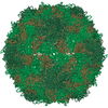
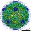
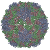
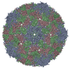
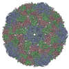
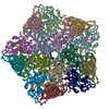
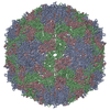
| タイトル | Cryo-EM structure of Poliovirus 135S particles | ||||||
 要素 要素 |
| ||||||
 キーワード キーワード |  VIRUS (ウイルス) / cell entry / single particle analysis (単粒子解析法) VIRUS (ウイルス) / cell entry / single particle analysis (単粒子解析法) | ||||||
| 機能・相同性 |  機能・相同性情報 機能・相同性情報symbiont-mediated suppression of host translation initiation / symbiont-mediated suppression of host cytoplasmic pattern recognition receptor signaling pathway via inhibition of RIG-I activity / symbiont-mediated suppression of host cytoplasmic pattern recognition receptor signaling pathway via inhibition of MDA-5 activity / symbiont-mediated suppression of host cytoplasmic pattern recognition receptor signaling pathway via inhibition of MAVS activity / ピコルナイン2A / symbiont-mediated suppression of host mRNA export from nucleus / ribonucleoside triphosphate phosphatase activity / symbiont genome entry into host cell via pore formation in plasma membrane / picornain 3C / T=pseudo3 icosahedral viral capsid ...symbiont-mediated suppression of host translation initiation / symbiont-mediated suppression of host cytoplasmic pattern recognition receptor signaling pathway via inhibition of RIG-I activity / symbiont-mediated suppression of host cytoplasmic pattern recognition receptor signaling pathway via inhibition of MDA-5 activity / symbiont-mediated suppression of host cytoplasmic pattern recognition receptor signaling pathway via inhibition of MAVS activity / ピコルナイン2A / symbiont-mediated suppression of host mRNA export from nucleus / ribonucleoside triphosphate phosphatase activity / symbiont genome entry into host cell via pore formation in plasma membrane / picornain 3C / T=pseudo3 icosahedral viral capsid / host cell cytoplasmic vesicle membrane / endocytosis involved in viral entry into host cell / : / nucleoside-triphosphate phosphatase / protein complex oligomerization / monoatomic ion channel activity / RNA helicase activity / induction by virus of host autophagy / RNA依存性RNAポリメラーゼ / symbiont-mediated suppression of host gene expression / viral RNA genome replication / cysteine-type endopeptidase activity / RNA-dependent RNA polymerase activity / DNA-templated transcription / host cell nucleus / virion attachment to host cell / structural molecule activity / タンパク質分解 / RNA binding / ATP binding / 生体膜 / metal ion binding類似検索 - 分子機能 | ||||||
| 生物種 |  Human poliovirus 1 (ポリオウイルス) Human poliovirus 1 (ポリオウイルス) | ||||||
| 手法 | 電子顕微鏡法 / 単粒子再構成法 / クライオ電子顕微鏡法 / 解像度: 5.5 Å | ||||||
 データ登録者 データ登録者 | Butan, C. / Fiman, D.J. / Hogle, J.M. | ||||||
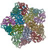
 引用 引用 | ジャーナル: J Virol / 年: 2014 タイトル: Cryo-electron microscopy reconstruction shows poliovirus 135S particles poised for membrane interaction and RNA release. 著者: Carmen Butan / David J Filman / James M Hogle /  要旨: During infection, binding of mature poliovirus to cell surface receptors induces an irreversible expansion of the capsid, to form an infectious cell-entry intermediate particle that sediments at 135S. ...During infection, binding of mature poliovirus to cell surface receptors induces an irreversible expansion of the capsid, to form an infectious cell-entry intermediate particle that sediments at 135S. In these expanded virions, the major capsid proteins (VP1 to VP3) adopt an altered icosahedral arrangement to open holes in the capsid at 2-fold and quasi-3-fold axes, and internal polypeptides VP4 and the N terminus of VP1, which can bind membranes, become externalized. Cryo-electron microscopy images for 117,330 particles were collected using Leginon and reconstructed using FREALIGN. Improved rigid-body positioning of major capsid proteins established reliably which polypeptide segments become disordered or rearranged. The virus-to-135S transition includes expansion of 4%, rearrangements of the GH loops of VP3 and VP1, and disordering of C-terminal extensions of VP1 and VP2. The N terminus of VP1 rearranges to become externalized near its quasi-3-fold exit, binds to rearranged GH loops of VP3 and VP1, and attaches to the top surface of VP2. These details improve our understanding of subsequent stages of infection, including endocytosis and RNA transfer into the cytoplasm. | ||||||
| 履歴 |
|
- 構造の表示
構造の表示
| ムービー |
ムービービューア |
|---|---|
| 構造ビューア | 分子: MolmilJmol/JSmol |
- ダウンロードとリンク
-ダウンロード
| PDBx/mmCIF形式 | 3j48.cif.gz | 38.4 KB | 表示 | PDBx/mmCIF形式 |
|---|---|---|---|---|
| PDB形式 | pdb3j48.ent.gz | 18.6 KB | 表示 | PDB形式 |
| PDBx/mmJSON形式 | 3j48.json.gz | ツリー表示 | PDBx/mmJSON形式 | |
| その他 |  その他のダウンロード その他のダウンロード |
-検証レポート
| アーカイブディレクトリ | https://data.pdbj.org/pub/pdb/validation_reports/j4/3j48ftp://data.pdbj.org/pub/pdb/validation_reports/j4/3j48 | HTTPS FTP |
|---|
-関連構造データ
-リンク
 PDBj
PDBj

- 集合体
集合体
| 登録構造単位 | 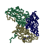
|
|---|---|
| 1 | x 60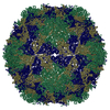
|
| 2 |
|
| 3 | x 5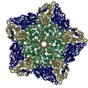
|
| 4 | x 6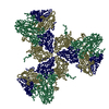
|
| 5 | 
|
| 対称性 | 点対称性: (シェーンフリース記号: I (正20面体型対称)) |
-要素
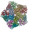
| #1: タンパク質 | 分子量: 33488.613 Da / 分子数: 1 / 断片: UNP residues 580-881 / 由来タイプ: 組換発現 由来: (組換発現) Human poliovirus 1 (ポリオウイルス)株: Mahoney / 細胞株 (発現宿主): HeLa / 発現宿主:  Homo sapiens (ヒト) / 参照: UniProt: P03300 Homo sapiens (ヒト) / 参照: UniProt: P03300 |
|---|---|
| #2: タンパク質 | 分子量: 30075.783 Da / 分子数: 1 / 断片: UNP residues 70-341 / 由来タイプ: 組換発現 由来: (組換発現) Human poliovirus 1 (ポリオウイルス)株: Mahoney / 細胞株 (発現宿主): HeLa / 発現宿主: Homo sapiens (ヒト) / 参照: UniProt: P03300 |
| #3: タンパク質 | 分子量: 26547.482 Da / 分子数: 1 / 断片: UNP residues 342-579 / 由来タイプ: 組換発現 由来: (組換発現) Human poliovirus 1 (ポリオウイルス)株: Mahoney / 細胞株 (発現宿主): HeLa / 発現宿主: Homo sapiens (ヒト) / 参照: UniProt: P03300 |
-実験情報
-実験
| 実験 | 手法: 電子顕微鏡法 |
|---|---|
| EM実験 | 試料の集合状態: PARTICLE / 3次元再構成法: 単粒子再構成法 |
- 試料調製
試料調製
| 構成要素 | 名称: Poliovirus 1 Mahoney 135S particle / タイプ: VIRUS 詳細: icosahedrally ordered capsid: 60 copies of VP1, VP2, VP3 | ||||||||||||
|---|---|---|---|---|---|---|---|---|---|---|---|---|---|
| 分子量 |
| ||||||||||||
| ウイルスについての詳細 | 中空か: NO / エンベロープを持つか: NO / ホストのカテゴリ: VERTEBRATES / 単離: STRAIN / タイプ: VIRION | ||||||||||||
| 天然宿主 | 生物種: Homo sapiens | ||||||||||||
| 緩衝液 | 名称: 20 mM HEPES, 2 mM CaCl2 / pH: 7.4 / 詳細: 20 mM HEPES, 2 mM CaCl2 | ||||||||||||
| 試料 | 濃度: 0.3 mg/ml / 包埋: NO / シャドウイング: NO / 染色: NO / 凍結: YES | ||||||||||||
| 試料支持 | 詳細: glow-discharged holey carbon-grids (200 mesh C-flat grids) | ||||||||||||
| 急速凍結 | 装置: HOMEMADE PLUNGER / 凍結剤: ETHANE / Temp: 90 K 詳細: Blotted manually in ambient atmosphere before plunging into ethane cooled by liquid nitrogen. 手法: Blotted manually before plunge freezing into liquid ethane |
- 電子顕微鏡撮影
| 実験機器 |  モデル: Tecnai F20 / 画像提供: FEI Company |
|---|---|
| 顕微鏡 | モデル: FEI TECNAI F20 / 日付: 2011年2月28日 |
| 電子銃 | 電子線源: FIELD EMISSION GUN / 加速電圧: 200 kV / 照射モード: FLOOD BEAM |
| 電子レンズ | モード: BRIGHT FIELDBright-field microscopy / 倍率(公称値): 62000 X / 最大 デフォーカス(公称値): 2000 nm / 最小 デフォーカス(公称値): 980 nm / Cs: 2 mm / 非点収差: Objective lens astigmatism was corrected |
| 試料ホルダ | 試料ホルダーモデル: GATAN LIQUID NITROGEN 資料ホルダタイプ: Side entry liquid nitrogen-cooled cryo specimen holder 温度: 90 K / 最高温度: 93 K / 最低温度: 90 K / 傾斜角・最大: 0 ° / 傾斜角・最小: 0 ° |
| 撮影 | 電子線照射量: 15 e/Å2 フィルム・検出器のモデル: TVIPS TEMCAM-F415 (4k x 4k) |
| 画像スキャン | デジタル画像の数: 1020 |
| 放射 | プロトコル: SINGLE WAVELENGTH / 単色(M)・ラウエ(L): M / 散乱光タイプ: x-ray |
| 放射波長 | 相対比: 1 |
- 解析
解析
| EMソフトウェア |
| ||||||||||||
|---|---|---|---|---|---|---|---|---|---|---|---|---|---|
| CTF補正 | 詳細: Each micrograph | ||||||||||||
| 対称性 | 点対称性: I (正20面体型対称) | ||||||||||||
| 3次元再構成 | 手法: FREALIGN randomized search / 解像度: 5.5 Å / 解像度の算出法: FSC 0.143 CUT-OFF / 粒子像の数: 117330 / ピクセルサイズ(公称値): 1.37 Å / ピクセルサイズ(実測値): 1.37 Å 詳細: Single particle details: The particles were selected using an automatic selection program (Single particle--Applied symmetry: I) 対称性のタイプ: POINT | ||||||||||||
| 原子モデル構築 | プロトコル: RIGID BODY FIT / 空間: RECIPROCAL Target criteria: mean amplitude-weighted cosine of the phase difference 詳細: METHOD--rigid body REFINEMENT PROTOCOL--rigid body DETAILS--Most of the model was docked, with specific areas of discrepancy fitted. The fitting was rigid body with flexible fitting or ...詳細: METHOD--rigid body REFINEMENT PROTOCOL--rigid body DETAILS--Most of the model was docked, with specific areas of discrepancy fitted. The fitting was rigid body with flexible fitting or deletion of selected polypeptide segments. Rigid bodies for VP1, VP2, VP3, and the VP3 beta tube were defined to include beta barrels and non-covalently attached polypeptides. Each rigid body was repeatedly fitted manually and then refined. Disordered polypeptide segments were removed. Several rearranged segments were included as approximate backbone traces and refined. | ||||||||||||
| 原子モデル構築 | 3D fitting-ID: 1 / Accession code: 1POV / Initial refinement model-ID: 1 / PDB-ID: 1POV / Source name: PDB / タイプ: experimental model
| ||||||||||||
| 精密化ステップ | サイクル: LAST
|