 ムービー
ムービー コントローラー


+ データを開く

- 基本情報
基本情報
| 登録情報 | データベース: EMDB / ID: EMD-1825 | |||||||||
|---|---|---|---|---|---|---|---|---|---|---|
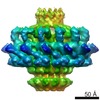
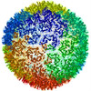
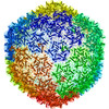
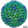
| タイトル | Structural basis for scaffolding-mediated assembly and maturation of a dsDNA virus | |||||||||
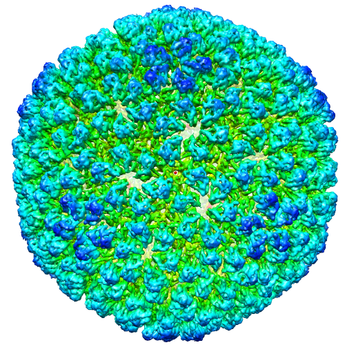
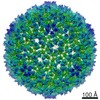
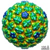
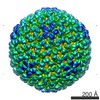
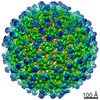
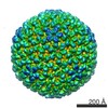
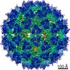
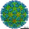
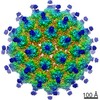
 マップデータ マップデータ | This is the icosahedral density map for bacteriophage P22 empty procapsid solved by cryo-EM at 7.0-Angstrom resolution | |||||||||
 試料 試料 |
| |||||||||
 キーワード キーワード |  bacteriophage (ファージ) / phage (ファージ) / P22 / procapsid (カプシド) / icosahedral reconstruction / dsDNA virus (DNAウイルス) bacteriophage (ファージ) / phage (ファージ) / P22 / procapsid (カプシド) / icosahedral reconstruction / dsDNA virus (DNAウイルス) | |||||||||
| 生物種 |  Enterobacteria phage P22 (ファージ) Enterobacteria phage P22 (ファージ) | |||||||||
| 手法 | 単粒子再構成法 / クライオ電子顕微鏡法 / 解像度: 7.0 Å | |||||||||
 データ登録者 データ登録者 | Chen D-H / Baker ML / Hryc CF / DiMaio F / Jakana J / Wu W / Dougherty M / Haase-Pettingell C / Schmid MF / Jiang W ...Chen D-H / Baker ML / Hryc CF / DiMaio F / Jakana J / Wu W / Dougherty M / Haase-Pettingell C / Schmid MF / Jiang W / Baker D / King JA / Chiu W | |||||||||
 引用 引用 | ジャーナル: Proc Natl Acad Sci U S A / 年: 2011 タイトル: Structural basis for scaffolding-mediated assembly and maturation of a dsDNA virus. 著者: Dong-Hua Chen / Matthew L Baker / Corey F Hryc / Frank DiMaio / Joanita Jakana / Weimin Wu / Matthew Dougherty / Cameron Haase-Pettingell / Michael F Schmid / Wen Jiang / David Baker / ...著者: Dong-Hua Chen / Matthew L Baker / Corey F Hryc / Frank DiMaio / Joanita Jakana / Weimin Wu / Matthew Dougherty / Cameron Haase-Pettingell / Michael F Schmid / Wen Jiang / David Baker / Jonathan A King / Wah Chiu /  要旨: Formation of many dsDNA viruses begins with the assembly of a procapsid, containing scaffolding proteins and a multisubunit portal but lacking DNA, which matures into an infectious virion. This ...Formation of many dsDNA viruses begins with the assembly of a procapsid, containing scaffolding proteins and a multisubunit portal but lacking DNA, which matures into an infectious virion. This process, conserved among dsDNA viruses such as herpes viruses and bacteriophages, is key to forming infectious virions. Bacteriophage P22 has served as a model system for this study in the past several decades. However, how capsid assembly is initiated, where and how scaffolding proteins bind to coat proteins in the procapsid, and the conformational changes upon capsid maturation still remain elusive. Here, we report Cα backbone models for the P22 procapsid and infectious virion derived from electron cryomicroscopy density maps determined at 3.8- and 4.0-Å resolution, respectively, and the first procapsid structure at subnanometer resolution without imposing symmetry. The procapsid structures show the scaffolding protein interacting electrostatically with the N terminus (N arm) of the coat protein through its C-terminal helix-loop-helix motif, as well as unexpected interactions between 10 scaffolding proteins and the 12-fold portal located at a unique vertex. These suggest a critical role for the scaffolding proteins both in initiating the capsid assembly at the portal vertex and propagating its growth on a T = 7 icosahedral lattice. Comparison of the procapsid and the virion backbone models reveals coordinated and complex conformational changes. These structural observations allow us to propose a more detailed molecular mechanism for the scaffolding-mediated capsid assembly initiation including portal incorporation, release of scaffolding proteins upon DNA packaging, and maturation into infectious virions. #1: ジャーナル: NAT.STRUCT.MOL.BIOL. / 年: 2003タイトル: Coat protein fold and maturation transition of bacteriophage P22 seen at subnanometer resolutions 著者: Jiang W / Li Z / Zhang Z / Baker ML / Prevelige PE Jr / Chiu W | |||||||||
| 履歴 |
|
- 構造の表示
構造の表示
| ムービー |
 ムービービューア ムービービューア |
|---|---|
| 構造ビューア | EMマップ: SurfViewMolmilJmol/JSmol |
| 添付画像 |
- ダウンロードとリンク
-EMDBアーカイブ
| マップデータ | emd_1825.map.gz | 276.4 MB | EMDBマップデータ形式 | |
|---|---|---|---|---|
| ヘッダ (付随情報) | emd-1825-v30.xmlemd-1825.xml | 11 KB 11 KB | 表示 表示 | EMDBヘッダ |
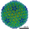
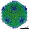
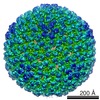
| 画像 |  EMD-1825.png EMD-1825.png | 421.7 KB | ||
| アーカイブディレクトリ |  http://ftp.pdbj.org/pub/emdb/structures/EMD-1825ftp://ftp.pdbj.org/pub/emdb/structures/EMD-1825 http://ftp.pdbj.org/pub/emdb/structures/EMD-1825ftp://ftp.pdbj.org/pub/emdb/structures/EMD-1825 | HTTPS FTP |
-関連構造データ
-リンク
| EMDBのページ | EMDB (EBI/PDBe) / EMDataResource |
|---|
-マップ
| ファイル | ダウンロード / ファイル: emd_1825.map.gz / 形式: CCP4 / 大きさ: 300.3 MB / タイプ: IMAGE STORED AS FLOATING POINT NUMBER (4 BYTES) | ||||||||||||||||||||||||||||||||||||||||||||||||||||||||||||||||||||
|---|---|---|---|---|---|---|---|---|---|---|---|---|---|---|---|---|---|---|---|---|---|---|---|---|---|---|---|---|---|---|---|---|---|---|---|---|---|---|---|---|---|---|---|---|---|---|---|---|---|---|---|---|---|---|---|---|---|---|---|---|---|---|---|---|---|---|---|---|---|
| 注釈 | This is the icosahedral density map for bacteriophage P22 empty procapsid solved by cryo-EM at 7.0-Angstrom resolution | ||||||||||||||||||||||||||||||||||||||||||||||||||||||||||||||||||||
| ボクセルのサイズ | X=Y=Z: 2.12 Å | ||||||||||||||||||||||||||||||||||||||||||||||||||||||||||||||||||||
| 密度 |
| ||||||||||||||||||||||||||||||||||||||||||||||||||||||||||||||||||||
| 対称性 | 空間群: 1 | ||||||||||||||||||||||||||||||||||||||||||||||||||||||||||||||||||||
| 詳細 | EMDB XML:
CCP4マップ ヘッダ情報:
| ||||||||||||||||||||||||||||||||||||||||||||||||||||||||||||||||||||
-添付データ
- 試料の構成要素
-全体 : Bacteriophage P22 empty procapsid
| 全体 | 名称: Bacteriophage P22 empty procapsid |
|---|---|
| 要素 |
|
-超分子 #1000: Bacteriophage P22 empty procapsid
| 超分子 | 名称: Bacteriophage P22 empty procapsid / タイプ: sample / ID: 1000 詳細: This sample is P22 empty procapsid from which the scaffolding proteins were removed by GuHCl. Number unique components: 1 |
|---|
-超分子 #1: Enterobacteria phage P22
| 超分子 | 名称: Enterobacteria phage P22 / タイプ: virus / ID: 1 / Name.synonym: P22 / NCBI-ID: 10754 / 生物種: Enterobacteria phage P22 / ウイルスタイプ: OTHER / ウイルス・単離状態: STRAIN / ウイルス・エンベロープ: No / ウイルス・中空状態: Yes / Syn species name: P22 |
|---|---|
| 宿主 | 生物種:  Salmonella enterica subsp. enterica serovar Typhimurium (サルモネラ菌) Salmonella enterica subsp. enterica serovar Typhimurium (サルモネラ菌)別称: BACTERIA(EUBACTERIA) |
| ウイルス殻 | Shell ID: 1 / 直径: 610 Å / T番号(三角分割数): 7 |
-実験情報
-構造解析
| 手法 | クライオ電子顕微鏡法 |
|---|---|
 解析 解析 | 単粒子再構成法 |
| 試料の集合状態 | particle |
-試料調製
| 濃度 | 1 mg/mL |
|---|---|
| 緩衝液 | pH: 7.6 / 詳細: 50 mM Tris pH 7.6, 25 mM NaCl, 2mM EDTA |
| グリッド | 詳細: 400 mesh copper grid |
| 凍結 | 凍結剤: ETHANE / チャンバー内湿度: 95 % / チャンバー内温度: 4.2 K / 装置: OTHER / 詳細: Vitrification instrument: Vitrobot / 手法: Blot for 2 seconds before plunging |
- 電子顕微鏡法
| 顕微鏡 | JEOL 3000SFF |
|---|---|
| 電子線 | 加速電圧: 300 kV / 電子線源: FIELD EMISSION GUN |
| 電子光学系 | 倍率(補正後): 60000 / 照射モード: FLOOD BEAM / 撮影モード: BRIGHT FIELDBright-field microscopy / Cs: 1.6 mm / 最大 デフォーカス(公称値): 4.0 µm / 最小 デフォーカス(公称値): 0.8 µm / 倍率(公称値): 60000 |
| 試料ステージ | 試料ホルダー: Top entry / 試料ホルダーモデル: JEOL |
| 温度 | 最低: 4.2 K / 最高: 4.2 K / 平均: 4.2 K |
| アライメント法 | Legacy - 非点収差: Objective lens astigmatism was corrected at 400,000 times magnification |
| 撮影 | カテゴリ: FILM / フィルム・検出器のモデル: KODAK SO-163 FILM / デジタル化 - スキャナー: NIKON COOLSCAN / デジタル化 - サンプリング間隔: 6.35 µm / 実像数: 570 / 平均電子線量: 25 e/Å2 / ビット/ピクセル: 8 |
-画像解析
| CTF補正 | 詳細: Each micrograph |
|---|---|
| 最終 角度割当 | 詳細: EMAN:az, alt, phi |
| 最終 再構成 | 想定した対称性 - 点群: I (正20面体型対称) / アルゴリズム: OTHER / 解像度のタイプ: BY AUTHOR / 解像度: 7.0 Å / 解像度の算出法: FSC 0.5 CUT-OFF ソフトウェア - 名称: Multi-Path Simulated Annealing Optimization algorithm 使用した粒子像数: 18200 |
| 詳細 | The particles were selected using an automatic selection program |
