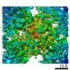
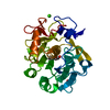
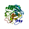
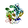
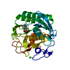
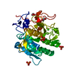
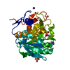
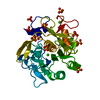
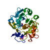
ジャーナル: J Struct Biol / 年: 2019 タイトル: Using focus ion beam to prepare crystal lamella for electron diffraction. 著者: Heng Zhou / Zhipu Luo / Xueming Li / 要旨: Electron diffraction provides a powerful tool to solve the structures of small protein crystals. However, strong interactions between the electrons and the materials limit the application of the ...Electron diffraction provides a powerful tool to solve the structures of small protein crystals. However, strong interactions between the electrons and the materials limit the application of the electron crystallographic method on large protein crystals with micrometer or larger sizes. Here, we used the focused ion beam (FIB) equipped on the scanning electron microscope (SEM) to mill a large crystal to thin lamella. The influences of the milling on the crystal lamella were observed and investigated, including radiation damage on the crystal surface during the FIB imaging, deformation of the thin crystal lamella, and variation in the diffraction intensities under electron radiation. These observations provide important information to optimize the FIB milling, and hence is important to obtain high-quality crystal samples for routine structure determination of protein crystals using the electron cryo-microscope.
∠α: 90 ° / ∠β: 90 ° / ∠γ: 90 ° / A: 69.31 Å / B: 69.31 Å / C: 104.12 Å / 空間群名: P43212 / 空間群番号: 96
CTF補正
タイプ: NONE
3次元再構成
解像度: 1.5 Å / 解像度の算出法: DIFFRACTION PATTERN/LAYERLINES / 対称性のタイプ: 3D CRYSTAL
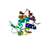
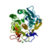
原子モデル構築
プロトコル: FLEXIBLE FIT / 空間: RECIPROCAL
精密化
解像度: 1.5→12.31 Å / Cor.coef. Fo:Fc: 0.956 / Cor.coef. Fo:Fc free: 0.938 / SU B: 2.195 / SU ML: 0.077 / 交差検証法: THROUGHOUT / σ(F): 0 / ESU R: 0.087 / ESU R Free: 0.09 / 立体化学のターゲット値: MAXIMUM LIKELIHOOD 詳細: HYDROGENS HAVE BEEN ADDED IN THE RIDING POSITIONS U VALUES : REFINED INDIVIDUALLY
Rfactor
反射数
%反射
Selection details
Rfree
0.2251
1893
5 %
RANDOM
Rwork
0.1868
-
-
-
obs
0.1888
35768
91.07 %
-
溶媒の処理
イオンプローブ半径: 0.8 Å / 減衰半径: 0.8 Å / VDWプローブ半径: 1.2 Å / 溶媒モデル: BABINET MODEL WITH MASK
 ムービー
ムービー コントローラー


 データを開く
 基本情報
基本情報 要素
要素 キーワード
キーワード 機能・相同性情報
 Parengyodontium album (菌類)
 データ登録者
 中国, 4件
  引用
引用 構造の表示
構造の表示 ダウンロードとリンク
 その他のダウンロード


 PDBj
PDBj

 集合体
集合体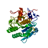

 分子量: 96.063 Da / 分子数: 1 / 由来タイプ: 合成 / 式: SO4
 分子量: 18.015 Da / 分子数: 230 / 由来タイプ: 天然 / 式: H2O
 試料調製
試料調製
 FIELD EMISSION GUN / 加速電圧: 200 kV / 照射モード: FLOOD BEAM
 解析
解析