Journal: J Struct Biol / Year: 2019 Title: Using focus ion beam to prepare crystal lamella for electron diffraction. Authors: Heng Zhou / Zhipu Luo / Xueming Li / Abstract: Electron diffraction provides a powerful tool to solve the structures of small protein crystals. However, strong interactions between the electrons and the materials limit the application of the ...Electron diffraction provides a powerful tool to solve the structures of small protein crystals. However, strong interactions between the electrons and the materials limit the application of the electron crystallographic method on large protein crystals with micrometer or larger sizes. Here, we used the focused ion beam (FIB) equipped on the scanning electron microscope (SEM) to mill a large crystal to thin lamella. The influences of the milling on the crystal lamella were observed and investigated, including radiation damage on the crystal surface during the FIB imaging, deformation of the thin crystal lamella, and variation in the diffraction intensities under electron radiation. These observations provide important information to optimize the FIB milling, and hence is important to obtain high-quality crystal samples for routine structure determination of protein crystals using the electron cryo-microscope.
Embedding applied: NO / Shadowing applied: NO / Staining applied: NO / Vitrification applied: YES
Vitrification
Cryogen name: ETHANE
-
Data collection
Experimental equipment
Model: Tecnai F20 / Image courtesy: FEI Company
Microscopy
Model: FEI TECNAI F20
Electron gun
Electron source: FIELD EMISSION GUN / Accelerating voltage: 200 kV / Illumination mode: FLOOD BEAM
Electron lens
Mode: DIFFRACTION
Image recording
Electron dose: 0.057 e/Å2 / Film or detector model: GATAN ULTRASCAN 4000 (4k x 4k)
EM diffraction
Camera length: 1200 mm
EM diffraction shell
Resolution: 1.7→1.76 Å / Fourier space coverage: 79.1 % / Multiplicity: 9.7 / Num. of structure factors: 1002 / Phase residual: 1 °
EM diffraction stats
Fourier space coverage: 89 % / High resolution: 1.7 Å / Num. of intensities measured: 121300 / Num. of structure factors: 11548 / Phase error: 0 ° / Phase residual: 1 ° / Phase error rejection criteria: 60 / Rmerge: 0.281 / Rsym: 0.281
Detector
Date: Feb 2, 2018
-
Processing
Software
Name
Version
Classification
NB
REFMAC
5.8.0222
refinement
PDB_EXTRACT
3.25
dataextraction
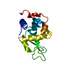
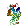
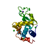
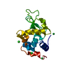
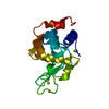
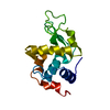
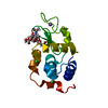
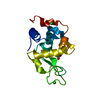
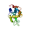
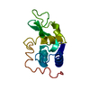
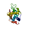
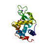
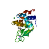
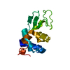
EM 3D crystal entity
∠α: 90 ° / ∠β: 90 ° / ∠γ: 90 ° / A: 78.99 Å / B: 78.99 Å / C: 37.89 Å / Space group name: P43212 / Space group num: 96
CTF correction
Type: NONE
3D reconstruction
Resolution: 1.73 Å / Resolution method: DIFFRACTION PATTERN/LAYERLINES / Symmetry type: 3D CRYSTAL
Atomic model building
Protocol: FLEXIBLE FIT / Space: RECIPROCAL
Refinement
Resolution: 1.73→14.67 Å / Cor.coef. Fo:Fc: 0.94 / Cor.coef. Fo:Fc free: 0.924 / SU B: 3.69 / SU ML: 0.113 / Cross valid method: THROUGHOUT / ESU R: 0.16 / ESU R Free: 0.147 Details: HYDROGENS HAVE BEEN ADDED IN THE RIDING POSITIONS U VALUES : REFINED INDIVIDUALLY
Rfactor
Num. reflection
% reflection
Selection details
Rfree
0.2575
550
4.8 %
RANDOM
Rwork
0.2203
-
-
-
obs
0.2221
10947
88.68 %
-
Solvent computation
Ion probe radii: 0.8 Å / Shrinkage radii: 0.8 Å / VDW probe radii: 1.2 Å
In the structure databanks used in Yorodumi, some data are registered as the other names, "COVID-19 virus" and "2019-nCoV". Here are the details of the virus and the list of structure data.
Jan 31, 2019. EMDB accession codes are about to change! (news from PDBe EMDB page)
EMDB accession codes are about to change! (news from PDBe EMDB page)
The allocation of 4 digits for EMDB accession codes will soon come to an end. Whilst these codes will remain in use, new EMDB accession codes will include an additional digit and will expand incrementally as the available range of codes is exhausted. The current 4-digit format prefixed with “EMD-” (i.e. EMD-XXXX) will advance to a 5-digit format (i.e. EMD-XXXXX), and so on. It is currently estimated that the 4-digit codes will be depleted around Spring 2019, at which point the 5-digit format will come into force.
The EM Navigator/Yorodumi systems omit the EMD- prefix.
Related info.:Q: What is EMD? / ID/Accession-code notation in Yorodumi/EM Navigator
Yorodumi is a browser for structure data from EMDB, PDB, SASBDB, etc.
This page is also the successor to EM Navigator detail page, and also detail information page/front-end page for Omokage search.
The word "yorodu" (or yorozu) is an old Japanese word meaning "ten thousand". "mi" (miru) is to see.
Related info.:EMDB / PDB / SASBDB / Comparison of 3 databanks / Yorodumi Search / Aug 31, 2016. New EM Navigator & Yorodumi / Yorodumi Papers / Jmol/JSmol / Function and homology information / Changes in new EM Navigator and Yorodumi
 Movie
Movie Controller

 Yorodumi
 Open data
 Basic information
 Components
 Keywords
 Function and homology information
 Authors
 China, 4items
  Citation
 Structure visualization
 Downloads & links
 Other downloads
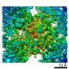


 PDBj
PDBj


 Assembly
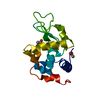

 Mass: 59.044 Da / Num. of mol.: 2 / Source method: obtained synthetically / Formula: C2H3O2
 Mass: 18.015 Da / Num. of mol.: 37 / Source method: isolated from a natural source / Formula: H2O
 Sample preparation
 FIELD EMISSION GUN / Accelerating voltage: 200 kV / Illumination mode: FLOOD BEAM
 Processing