Mass: 18.015 Da / Num. of mol.: 108 / Source method: isolated from a natural source / Formula: H2O
-
Details
Has protein modification
Y
-
Experimental details
-
Experiment
Experiment
Method: X-RAY DIFFRACTION / Number of used crystals: 1
-
Sample preparation
Crystal
Density Matthews: 2.08 Å3/Da / Density % sol: 40.84 %
Crystal grow
Temperature: 295 K / Method: batch mode / pH: 4.7 Details: Hen Egg White Lysozyme (49mg, ie 3.2mM), cisplatin (3mg, ie 10mM), 462.5 microlitres of 0.04 M sodium acetate, 462.5 microlitres of 10% sodium chloride, and with 7.5% DMSO (75 microlitres ie ...Details: Hen Egg White Lysozyme (49mg, ie 3.2mM), cisplatin (3mg, ie 10mM), 462.5 microlitres of 0.04 M sodium acetate, 462.5 microlitres of 10% sodium chloride, and with 7.5% DMSO (75 microlitres ie 1mM), pH 4.7, batch crystallisation method and room temperature (295K). The crystallisation pot was kept for five years.
Resolution: 1→27.386 Å / SU ML: 0.14 / SU R Cruickshank DPI: 0.027 / Cross valid method: FREE R-VALUE / σ(F): 1.35 / Phase error: 31.93 Details: Both Phenix_Refine (Afonine et al 2012) and CCP4 Refmac (Murshudov et al 1997) were used for the model refinement, taking advantage of the advantages of each. COOT (Emsley and Cowtan 2004) ...Details: Both Phenix_Refine (Afonine et al 2012) and CCP4 Refmac (Murshudov et al 1997) were used for the model refinement, taking advantage of the advantages of each. COOT (Emsley and Cowtan 2004) was used to inspect the molecular model and the electron density maps.
Rfactor
Num. reflection
% reflection
Selection details
Rfree
0.2028
3013
5.23 %
Random selection
Rwork
0.1758
-
-
-
obs
0.1772
57620
87.8 %
-
Solvent computation
Shrinkage radii: 0.9 Å / VDW probe radii: 1.11 Å
Displacement parameters
Biso mean: 19.4 Å2
Refinement step
Cycle: LAST / Resolution: 1→27.386 Å
Protein
Nucleic acid
Ligand
Solvent
Total
Num. atoms
1001
0
15
108
1124
Refine LS restraints
Refine-ID
Type
Dev ideal
Number
X-RAY DIFFRACTION
f_bond_d
0.012
1142
X-RAY DIFFRACTION
f_angle_d
1.274
1550
X-RAY DIFFRACTION
f_dihedral_angle_d
12.413
423
X-RAY DIFFRACTION
f_chiral_restr
0.078
156
X-RAY DIFFRACTION
f_plane_restr
0.006
211
LS refinement shell
Resolution (Å)
Rfactor Rfree
Num. reflection Rfree
Rfactor Rwork
Num. reflection Rwork
Refine-ID
% reflection obs (%)
1-1.0156
0.3638
79
0.3659
1660
X-RAY DIFFRACTION
59
1.0156-1.0323
0.3956
114
0.3661
2034
X-RAY DIFFRACTION
73
1.0323-1.0501
0.4059
136
0.3537
2197
X-RAY DIFFRACTION
79
1.0501-1.0692
0.3699
115
0.3564
2382
X-RAY DIFFRACTION
85
1.0692-1.0897
0.4261
135
0.3474
2422
X-RAY DIFFRACTION
87
1.0897-1.112
0.3551
146
0.2886
2517
X-RAY DIFFRACTION
91
1.112-1.1362
0.3122
133
0.2751
2607
X-RAY DIFFRACTION
93
1.1362-1.1626
0.2697
168
0.2404
2590
X-RAY DIFFRACTION
94
1.1626-1.1917
0.2306
131
0.2137
2648
X-RAY DIFFRACTION
94
1.1917-1.2239
0.2593
152
0.2007
2633
X-RAY DIFFRACTION
94
1.2239-1.2599
0.216
141
0.1906
2676
X-RAY DIFFRACTION
94
1.2599-1.3006
0.233
124
0.1823
2621
X-RAY DIFFRACTION
94
1.3006-1.347
0.2256
152
0.18
2622
X-RAY DIFFRACTION
93
1.347-1.401
0.1989
158
0.1629
2576
X-RAY DIFFRACTION
92
1.401-1.4647
0.1744
161
0.1559
2618
X-RAY DIFFRACTION
93
1.4647-1.542
0.1846
132
0.1544
2599
X-RAY DIFFRACTION
92
1.542-1.6386
0.1597
136
0.1496
2564
X-RAY DIFFRACTION
91
1.6386-1.765
0.1683
152
0.1484
2544
X-RAY DIFFRACTION
90
1.765-1.9426
0.1833
139
0.149
2549
X-RAY DIFFRACTION
89
1.9426-2.2236
0.1864
136
0.1528
2533
X-RAY DIFFRACTION
88
2.2236-2.8011
0.1603
122
0.1668
2500
X-RAY DIFFRACTION
85
2.8011-27.3962
0.2055
151
0.1707
2515
X-RAY DIFFRACTION
82
+
About Yorodumi
-
News
-
Feb 9, 2022. New format data for meta-information of EMDB entries
New format data for meta-information of EMDB entries
Version 3 of the EMDB header file is now the official format.
The previous official version 1.9 will be removed from the archive.
In the structure databanks used in Yorodumi, some data are registered as the other names, "COVID-19 virus" and "2019-nCoV". Here are the details of the virus and the list of structure data.
Jan 31, 2019. EMDB accession codes are about to change! (news from PDBe EMDB page)
EMDB accession codes are about to change! (news from PDBe EMDB page)
The allocation of 4 digits for EMDB accession codes will soon come to an end. Whilst these codes will remain in use, new EMDB accession codes will include an additional digit and will expand incrementally as the available range of codes is exhausted. The current 4-digit format prefixed with “EMD-” (i.e. EMD-XXXX) will advance to a 5-digit format (i.e. EMD-XXXXX), and so on. It is currently estimated that the 4-digit codes will be depleted around Spring 2019, at which point the 5-digit format will come into force.
The EM Navigator/Yorodumi systems omit the EMD- prefix.
Related info.:Q: What is EMD? / ID/Accession-code notation in Yorodumi/EM Navigator
Yorodumi is a browser for structure data from EMDB, PDB, SASBDB, etc.
This page is also the successor to EM Navigator detail page, and also detail information page/front-end page for Omokage search.
The word "yorodu" (or yorozu) is an old Japanese word meaning "ten thousand". "mi" (miru) is to see.
Related info.:EMDB / PDB / SASBDB / Comparison of 3 databanks / Yorodumi Search / Aug 31, 2016. New EM Navigator & Yorodumi / Yorodumi Papers / Jmol/JSmol / Function and homology information / Changes in new EM Navigator and Yorodumi
 Movie
Movie Controller

 Yorodumi
 Open data
 Basic information
 Components
 Keywords
 Function and homology information
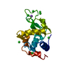
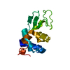
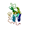
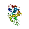
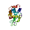
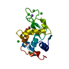
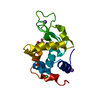
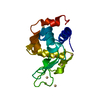
 X-RAY DIFFRACTION /
  Authors
 Citation
 Structure visualization
 Downloads & links
 Other downloads


 PDBj
PDBj


 Assembly


 Mass: 78.133 Da / Num. of mol.: 1 / Source method: obtained synthetically / Formula: C2H6OS / Comment: DMSO, precipitant*YM
 Mass: 35.453 Da / Num. of mol.: 5 / Source method: obtained synthetically / Formula: Cl
 Mass: 22.990 Da / Num. of mol.: 1 / Source method: obtained synthetically / Formula: Na
 Mass: 195.078 Da / Num. of mol.: 3 / Source method: obtained synthetically / Formula: Pt
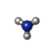
 Mass: 17.031 Da / Num. of mol.: 2 / Source method: obtained synthetically / Formula: NH3
 Sample preparation
 / Beamline: I04 / Wavelength: 0.92819 Å
 Processing