 Movie
Movie Controller

[English] 日本語
 Yorodumi
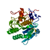
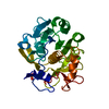
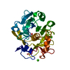
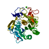
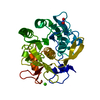
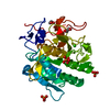
- PDB-6qxv: Pink beam serial crystallography: Proteinase K, 1 us exposure, 15... -
+ Open data

- Basic information
| Entry | Database: PDB / ID: 6qxv | ||||||||||||||||||
|---|---|---|---|---|---|---|---|---|---|---|---|---|---|---|---|---|---|---|---|
| Title | Pink beam serial crystallography: Proteinase K, 1 us exposure, 1585 patterns merged (2 chips) | ||||||||||||||||||
 Components Components | Proteinase K | ||||||||||||||||||
 Keywords Keywords | HYDROLASE / pink beam / fixed-target / serial crystallography / proteinase K | ||||||||||||||||||
| Function / homology |  Function and homology information Function and homology informationpeptidase K / serine-type endopeptidase activity / proteolysis / extracellular region / metal ion binding Similarity search - Function | ||||||||||||||||||
| Biological species |  Parengyodontium album (fungus) Parengyodontium album (fungus) | ||||||||||||||||||
| Method |  X-RAY DIFFRACTION / SYNCHROTRON / MOLECULAR REPLACEMENT / Resolution: 1.94 Å X-RAY DIFFRACTION / SYNCHROTRON / MOLECULAR REPLACEMENT / Resolution: 1.94 Å | ||||||||||||||||||
 Authors Authors | Tolstikova, A. / Oberthuer, D. / Meents, A. | ||||||||||||||||||
| Funding support |  Germany, 5items Germany, 5items
| ||||||||||||||||||
 Citation Citation | Journal: Iucrj / Year: 2019 Title: 1 kHz fixed-target serial crystallography using a multilayer monochromator and an integrating pixel detector. Authors: Tolstikova, A. / Levantino, M. / Yefanov, O. / Hennicke, V. / Fischer, P. / Meyer, J. / Mozzanica, A. / Redford, S. / Crosas, E. / Opara, N.L. / Barthelmess, M. / Lieske, J. / Oberthuer, D. ...Authors: Tolstikova, A. / Levantino, M. / Yefanov, O. / Hennicke, V. / Fischer, P. / Meyer, J. / Mozzanica, A. / Redford, S. / Crosas, E. / Opara, N.L. / Barthelmess, M. / Lieske, J. / Oberthuer, D. / Wator, E. / Mohacsi, I. / Wulff, M. / Schmitt, B. / Chapman, H.N. / Meents, A. | ||||||||||||||||||
| History |
|
- Structure visualization
| Structure viewer | Molecule: MolmilJmol/JSmol |
|---|
- Downloads & links
-Download
| PDBx/mmCIF format | 6qxv.cif.gz | 122.5 KB | Display | PDBx/mmCIF format |
|---|---|---|---|---|
| PDB format | pdb6qxv.ent.gz | 96.2 KB | Display | PDB format |
| PDBx/mmJSON format | 6qxv.json.gz | Tree view | PDBx/mmJSON format | |
| Others |  Other downloads Other downloads |
-Validation report
| Arichive directory | https://data.pdbj.org/pub/pdb/validation_reports/qx/6qxvftp://data.pdbj.org/pub/pdb/validation_reports/qx/6qxv | HTTPS FTP |
|---|
-Related structure data
| Related structure data |  6qxwC  6qxxC 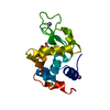 6qxyC  6qy0C  6qy1C  6qy2C  6qy4C  6qy5C  5kxvS S: Starting model for refinement C: citing same article ( |
|---|---|
| Similar structure data |


-Links
 PDBj
PDBj

- Assembly
| Deposited unit | 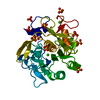
| |||||||||
|---|---|---|---|---|---|---|---|---|---|---|
| 1 |
| |||||||||
| Unit cell |
| |||||||||
| Components on special symmetry positions |
|
-Components
| #1: Protein | Mass: 28958.791 Da / Num. of mol.: 1 Source method: isolated from a genetically manipulated source Details: S312D mutation / Source: (gene. exp.) Parengyodontium album (fungus) / Gene: PROK / Production host: Engyodontium album (fungus) / References: UniProt: P06873, peptidase K | ||||||||
|---|---|---|---|---|---|---|---|---|---|
| #2: Chemical |   Mass: 40.078 Da / Num. of mol.: 2 / Source method: obtained synthetically / Formula: Ca Mass: 40.078 Da / Num. of mol.: 2 / Source method: obtained synthetically / Formula: Ca#3: Chemical | ChemComp-SO4 /   Mass: 96.063 Da / Num. of mol.: 11 / Source method: obtained synthetically / Formula: SO4 Mass: 96.063 Da / Num. of mol.: 11 / Source method: obtained synthetically / Formula: SO4#4: Chemical |   Mass: 35.453 Da / Num. of mol.: 3 / Source method: obtained synthetically / Formula: Cl Mass: 35.453 Da / Num. of mol.: 3 / Source method: obtained synthetically / Formula: Cl#5: Water | ChemComp-HOH / |  Mass: 18.015 Da / Num. of mol.: 187 / Source method: isolated from a natural source / Formula: H2O Mass: 18.015 Da / Num. of mol.: 187 / Source method: isolated from a natural source / Formula: H2OHas protein modification | Y | |
-Experimental details
-Experiment
| Experiment | Method: X-RAY DIFFRACTION / Number of used crystals: 1 |
|---|
- Sample preparation
| Crystal | Density Matthews: 2.12 Å3/Da / Density % sol: 42.1 % |
|---|---|
| Crystal grow | Temperature: 298 K / Method: vapor diffusion, sitting drop / pH: 6.6 Details: 0.1M CHC buffer pH 6.6, 0.7M ammonium sulphate and 10mM calcium chloride |
-Data collection
| Diffraction | Mean temperature: 298 K / Serial crystal experiment: Y | |||||||||
|---|---|---|---|---|---|---|---|---|---|---|
| Diffraction source | Source: SYNCHROTRON / Site: ESRF  / Beamline: ID09 / Wavelength: 0.80580-0.8262 / Beamline: ID09 / Wavelength: 0.80580-0.8262 | |||||||||
| Detector | Type: PSI JUNGFRAU 1M / Detector: PIXEL / Date: Jul 15, 2017 | |||||||||
| Radiation | Protocol: LAUE / Monochromatic (M) / Laue (L): L / Scattering type: x-ray | |||||||||
| Radiation wavelength |
| |||||||||
| Reflection | Resolution: 1.94→21.7 Å / Num. obs: 18492 / % possible obs: 96.25 % / Redundancy: 23.1 % / Biso Wilson estimate: 22.79 Å2 / CC1/2: 0.8763 / Net I/σ(I): 4.6 | |||||||||
| Reflection shell | Resolution: 1.94→2.009 Å / CC1/2: 0.7166 | |||||||||
| Serial crystallography sample delivery | Description: Roadrunner II / Method: fixed target | |||||||||
| Serial crystallography sample delivery fixed target | Description: Roadrunner II |
- Processing
| Software |
| ||||||||||||||||||||||||||||||||||||||||||||||||||||||||||||||||||||||||||||||||||||||||||||||||||
|---|---|---|---|---|---|---|---|---|---|---|---|---|---|---|---|---|---|---|---|---|---|---|---|---|---|---|---|---|---|---|---|---|---|---|---|---|---|---|---|---|---|---|---|---|---|---|---|---|---|---|---|---|---|---|---|---|---|---|---|---|---|---|---|---|---|---|---|---|---|---|---|---|---|---|---|---|---|---|---|---|---|---|---|---|---|---|---|---|---|---|---|---|---|---|---|---|---|---|---|
| Refinement | Method to determine structure: MOLECULAR REPLACEMENT Starting model: 5KXV Resolution: 1.94→21.7 Å / SU ML: 0.2 / Cross valid method: FREE R-VALUE / σ(F): 1.34 / Phase error: 22.1
| ||||||||||||||||||||||||||||||||||||||||||||||||||||||||||||||||||||||||||||||||||||||||||||||||||
| Solvent computation | Shrinkage radii: 0.9 Å / VDW probe radii: 1.11 Å | ||||||||||||||||||||||||||||||||||||||||||||||||||||||||||||||||||||||||||||||||||||||||||||||||||
| Displacement parameters | Biso max: 147.6 Å2 / Biso mean: 26.0448 Å2 / Biso min: 8 Å2 | ||||||||||||||||||||||||||||||||||||||||||||||||||||||||||||||||||||||||||||||||||||||||||||||||||
| Refinement step | Cycle: final / Resolution: 1.94→21.7 Å
| ||||||||||||||||||||||||||||||||||||||||||||||||||||||||||||||||||||||||||||||||||||||||||||||||||
| LS refinement shell | Refine-ID: X-RAY DIFFRACTION / Rfactor Rfree error: 0 / Total num. of bins used: 13
|
