Group: Data collection / Derived calculations ...Data collection / Derived calculations / Other / Source and taxonomy / Structure summary / Version format compliance
Monochromator: SI(111) CCM / Protocol: SINGLE WAVELENGTH / Monochromatic (M) / Laue (L): M / Scattering type: x-ray
Radiation wavelength
Wavelength: 0.8551 Å / Relative weight: 1
Reflection
Resolution: 0.95→20 Å / Num. obs: 1875402 / % possible obs: 98.7 % / Observed criterion σ(I): 2 / Redundancy: 6.3 % / Rmerge(I) obs: 0.05 / Net I/σ(I): 30.5
Reflection shell
Resolution: 0.94→0.95 Å / Redundancy: 2.2 % / Rmerge(I) obs: 0.3 / Mean I/σ(I) obs: 2 / % possible all: 85.6
-
Processing
Software
Name
Version
Classification
REFMAC
5.2.0019
refinement
HKL-2000
datareduction
HKL-2000
datascaling
SHELX
phasing
Refinement
Method to determine structure: SAD Starting model: NONE Resolution: 0.94→20 Å / Cor.coef. Fo:Fc: 0.986 / Cor.coef. Fo:Fc free: 0.982 / SU B: 0.259 / SU ML: 0.007 / Cross valid method: THROUGHOUT / ESU R: 0.013 / ESU R Free: 0.014 / Stereochemistry target values: MAXIMUM LIKELIHOOD / Details: HYDROGENS HAVE BEEN ADDED IN THE RIDING POSITIONS
Rfactor
Num. reflection
% reflection
Selection details
Rfree
0.103
7746
5 %
RANDOM
Rwork
0.09
-
-
-
obs
0.091
146726
99.1 %
-
Solvent computation
Ion probe radii: 0.8 Å / Shrinkage radii: 0.8 Å / VDW probe radii: 1.2 Å / Solvent model: BABINET MODEL WITH MASK
 Movie
Movie Controller

 Yorodumi
 Open data
 Basic information
 Components
 Keywords
 Function and homology information
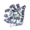
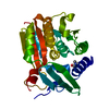
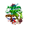
 ENGYODONTIUM ALBUM (fungus)
 X-RAY DIFFRACTION /
  Authors
 Citation
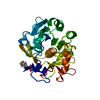
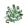
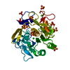
 Structure visualization
 Downloads & links
 Other downloads
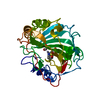


 PDBj
PDBj

 Assembly
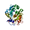

 Mass: 40.078 Da / Num. of mol.: 1 / Source method: obtained synthetically / Formula: Ca
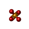
 Mass: 142.958 Da / Num. of mol.: 1 / Source method: obtained synthetically / Formula: O4Se
 Mass: 18.015 Da / Num. of mol.: 538 / Source method: isolated from a natural source / Formula: H2O
 Sample preparation
 / Beamline: X6A / Wavelength: 0.8551
  Processing