 ムービー
ムービー コントローラー


+ データを開く

- 基本情報
基本情報
| 登録情報 | データベース: PDB / ID: 6gyb | ||||||||||||
|---|---|---|---|---|---|---|---|---|---|---|---|---|---|
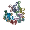
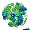
| タイトル | Cryo-EM structure of the bacteria-killing type IV secretion system core complex from Xanthomonas citri | ||||||||||||
 要素 要素 |
| ||||||||||||
 キーワード キーワード |  MEMBRANE PROTEIN (膜タンパク質) / core complex / bacterial killing / protein transport / bacterial Type IV Secretion System (分泌) MEMBRANE PROTEIN (膜タンパク質) / core complex / bacterial killing / protein transport / bacterial Type IV Secretion System (分泌) | ||||||||||||
| 機能・相同性 |  機能・相同性情報 機能・相同性情報 | ||||||||||||
| 生物種 |  Xanthomonas axonopodis pv. citri (バクテリア) Xanthomonas axonopodis pv. citri (バクテリア) | ||||||||||||
| 手法 | 電子顕微鏡法 / 単粒子再構成法 / クライオ電子顕微鏡法 / 解像度: 3.28 Å | ||||||||||||
 データ登録者 データ登録者 | Sgro, G.G. / Costa, T.R.D. / Farah, C.S. / Waksman, G. | ||||||||||||
| 資金援助 |  英国, 英国,  ブラジル, 3件 ブラジル, 3件
| ||||||||||||
 引用 引用 | ジャーナル: Nat Microbiol / 年: 2018 タイトル: Cryo-EM structure of the bacteria-killing type IV secretion system core complex from Xanthomonas citri. 著者: Germán G Sgro / Tiago R D Costa / William Cenens / Diorge P Souza / Alexandre Cassago / Luciana Coutinho de Oliveira / Roberto K Salinas / Rodrigo V Portugal / Chuck S Farah / Gabriel Waksman /  要旨: Type IV secretion (T4S) systems form the most common and versatile class of secretion systems in bacteria, capable of injecting both proteins and DNAs into host cells. T4S systems are typically ...Type IV secretion (T4S) systems form the most common and versatile class of secretion systems in bacteria, capable of injecting both proteins and DNAs into host cells. T4S systems are typically composed of 12 components that form 2 major assemblies: the inner membrane complex embedded in the inner membrane and the core complex embedded in both the inner and outer membranes. Here we present the 3.3 Å-resolution cryo-electron microscopy model of the T4S system core complex from Xanthomonas citri, a phytopathogen that utilizes this system to kill bacterial competitors. An extensive mutational investigation was performed to probe the vast network of protein-protein interactions in this 1.13-MDa assembly. This structure expands our knowledge of the molecular details of T4S system organization, assembly and evolution. | ||||||||||||
| 履歴 |
|
- 構造の表示
構造の表示
| ムービー |
ムービービューア |
|---|---|
| 構造ビューア | 分子: MolmilJmol/JSmol |
- ダウンロードとリンク
-ダウンロード
| PDBx/mmCIF形式 | 6gyb.cif.gz | 1.3 MB | 表示 | PDBx/mmCIF形式 |
|---|---|---|---|---|
| PDB形式 | pdb6gyb.ent.gz | 1.1 MB | 表示 | PDB形式 |
| PDBx/mmJSON形式 | 6gyb.json.gz | ツリー表示 | PDBx/mmJSON形式 | |
| その他 |  その他のダウンロード その他のダウンロード |
-検証レポート
| アーカイブディレクトリ | https://data.pdbj.org/pub/pdb/validation_reports/gy/6gybftp://data.pdbj.org/pub/pdb/validation_reports/gy/6gyb | HTTPS FTP |
|---|
-関連構造データ
| 関連構造データ | 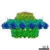 0089MC M: このデータのモデリングに利用したマップデータ C: 同じ文献を引用 ( |
|---|---|
| 類似構造データ | |
| その他のデータベース |
|


-リンク
 PDBj
PDBj- 集合体
集合体
| 登録構造単位 | 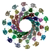
|
|---|---|
| 1 |
|
-要素
| #1: タンパク質 | 分子量: 14762.795 Da / 分子数: 14 / 由来タイプ: 組換発現 由来: (組換発現) Xanthomonas axonopodis pv. citri (strain 306) (バクテリア)遺伝子: XAC2622 / 発現宿主: Escherichia coli BL21(DE3) (大腸菌) / 参照: UniProt: Q8PJB3#2: タンパク質 | 分子量: 29359.385 Da / 分子数: 14 / 由来タイプ: 組換発現 由来: (組換発現) Xanthomonas axonopodis pv. citri (strain 306) (バクテリア)株: 306 / 遺伝子: virB9, XAC2620 / 発現宿主: Escherichia coli BL21(DE3) (大腸菌) / 参照: UniProt: Q8PJB5#3: タンパク質 | 分子量: 43392.469 Da / 分子数: 14 / 由来タイプ: 組換発現 由来: (組換発現) Xanthomonas axonopodis pv. citri (strain 306) (バクテリア)株: 306 / 遺伝子: virB10, XAC2619 / 発現宿主: Escherichia coli BL21(DE3) (大腸菌) / 参照: UniProt: Q8PJB6 |
|---|
-実験情報
-実験
| 実験 | 手法: 電子顕微鏡法 |
|---|---|
| EM実験 | 試料の集合状態: PARTICLE / 3次元再構成法: 単粒子再構成法 |
- 試料調製
試料調製
| 構成要素 | 名称: Core complex of a bacterial killing type IV secretion system from Xanthomonas分泌 タイプ: COMPLEX 詳細: Fourteen copies of each of the following three subunits: VirB7, VirB9 and VirB10 Entity ID: all / 由来: RECOMBINANT | ||||||||||||||||||||
|---|---|---|---|---|---|---|---|---|---|---|---|---|---|---|---|---|---|---|---|---|---|
| 分子量 | 実験値: NO | ||||||||||||||||||||
| 由来(天然) | 生物種: Xanthomonas axonopodis pv. citri str. 306 (バクテリア) | ||||||||||||||||||||
| 由来(組換発現) | 生物種: Escherichia coli BL21(DE3) (大腸菌) | ||||||||||||||||||||
| 緩衝液 | pH: 8 | ||||||||||||||||||||
| 緩衝液成分 |
| ||||||||||||||||||||
| 試料 | 濃度: 0.3 mg/ml / 包埋: NO / シャドウイング: NO / 染色: NO / 凍結: YES | ||||||||||||||||||||
| 急速凍結 | 装置: FEI VITROBOT MARK IV / 凍結剤: ETHANE / 湿度: 100 % / 凍結前の試料温度: 295.2 K 詳細: Blot for 4.5 seconds after 30 seconds of incubation. |
- 電子顕微鏡撮影
| 実験機器 |  モデル: Titan Krios / 画像提供: FEI Company |
|---|---|
| 顕微鏡 | モデル: FEI TITAN KRIOS |
| 電子銃 | 電子線源: FIELD EMISSION GUN / 加速電圧: 300 kV / 照射モード: FLOOD BEAM |
| 電子レンズ | モード: BRIGHT FIELDBright-field microscopy |
| 撮影 | 平均露光時間: 12 sec. / 電子線照射量: 60 e/Å2 / 検出モード: SUPER-RESOLUTION フィルム・検出器のモデル: GATAN K2 QUANTUM (4k x 4k) 撮影したグリッド数: 1 / 実像数: 1469 |
| 画像スキャン | 動画フレーム数/画像: 40 |
- 解析
解析
| ソフトウェア | 名称: PHENIX / バージョン: 1.12_2829: / 分類: 精密化 | ||||||||||||||||||||||||||||||||||||||||||||
|---|---|---|---|---|---|---|---|---|---|---|---|---|---|---|---|---|---|---|---|---|---|---|---|---|---|---|---|---|---|---|---|---|---|---|---|---|---|---|---|---|---|---|---|---|---|
| EMソフトウェア |
| ||||||||||||||||||||||||||||||||||||||||||||
| CTF補正 | タイプ: PHASE FLIPPING AND AMPLITUDE CORRECTION | ||||||||||||||||||||||||||||||||||||||||||||
| 粒子像の選択 | 選択した粒子像数: 185079 | ||||||||||||||||||||||||||||||||||||||||||||
| 対称性 | 点対称性: C14 (14回回転対称) | ||||||||||||||||||||||||||||||||||||||||||||
| 3次元再構成 | 解像度: 3.28 Å / 解像度の算出法: FSC 0.143 CUT-OFF / 粒子像の数: 142306 / アルゴリズム: BACK PROJECTION / クラス平均像の数: 1 / 対称性のタイプ: POINT | ||||||||||||||||||||||||||||||||||||||||||||
| 原子モデル構築 | B value: 138 / 空間: REAL / Target criteria: Cross-correlation coefficient 詳細: The electron density was clearly interpretable, which allowed us to build a de novo structural model. This process began by fitting the crystallographic model of the X. citri VirB7 C-terminal ...詳細: The electron density was clearly interpretable, which allowed us to build a de novo structural model. This process began by fitting the crystallographic model of the X. citri VirB7 C-terminal N0 domain (PDB:3OV5) and the NMR model of the X. citri VirB9CTD-VirB7NTD complex (PDB:2N01) in order to identify the map with the correct handedness. Models were positioned using Fit in map tool in Chimera, and saved relative to the map. Using these as starting points, we were able to manually build the rest of the model for VirB7 and VirB9CTD, and the de novo models for VirB10CTD, VirB10NTD_150-161 and VirB9NTD using Coot. In this manner, we obtained a combined model for a single VirB7-VirB9-VirB10 heterotrimer unit, which was submitted to iterative rounds of real space refinement and building using PHENIX and Coot software, respectively. Thirteen more copies of the refined heterotrimer were then fit into the density map using Chimera and new rounds of real space refinement (now using NCS for the 42 chains contained in the structure) and building using PHENIX and Coot, respectively, were executed until we obtained good parameters for Ramachandran plot and MolProbity. Chimera and PyMol were used for map and model visualization and figure production. | ||||||||||||||||||||||||||||||||||||||||||||
| 拘束条件 |
|
