 Movie
Movie Controller

[English] 日本語
 Yorodumi
- EMDB-0089: Cryo-EM structure of the bacteria-killing type IV secretion syste... -
+ Open data

- Basic information
| Entry |  | ||||||||||||
|---|---|---|---|---|---|---|---|---|---|---|---|---|---|
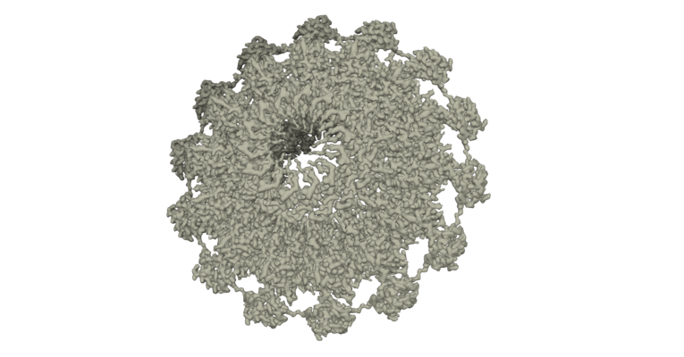
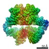
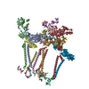
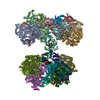
| Title | Cryo-EM structure of the bacteria-killing type IV secretion system core complex from Xanthomonas citri | ||||||||||||
 Map data Map data | |||||||||||||
 Sample Sample |
| ||||||||||||
 Keywords Keywords | core complex / bacterial killing / protein transport / bacterial Type IV Secretion System / MEMBRANE PROTEIN | ||||||||||||
| Function / homology |  Function and homology information Function and homology information | ||||||||||||
| Biological species |  Xanthomonas axonopodis pv. citri str. 306 (bacteria) / Xanthomonas axonopodis pv. citri (strain 306) (bacteria) Xanthomonas axonopodis pv. citri str. 306 (bacteria) / Xanthomonas axonopodis pv. citri (strain 306) (bacteria) | ||||||||||||
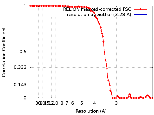
| Method | single particle reconstruction / cryo EM / Resolution: 3.28 Å | ||||||||||||
 Authors Authors | Costa TRD / Sgro GG / Farah CS / Waksman G | ||||||||||||
| Funding support |  United Kingdom, United Kingdom,  Brazil, 3 items Brazil, 3 items
| ||||||||||||
 Citation Citation | Journal: Nat Microbiol / Year: 2018 Title: Cryo-EM structure of the bacteria-killing type IV secretion system core complex from Xanthomonas citri. Authors: Germán G Sgro / Tiago R D Costa / William Cenens / Diorge P Souza / Alexandre Cassago / Luciana Coutinho de Oliveira / Roberto K Salinas / Rodrigo V Portugal / Chuck S Farah / Gabriel Waksman /  Abstract: Type IV secretion (T4S) systems form the most common and versatile class of secretion systems in bacteria, capable of injecting both proteins and DNAs into host cells. T4S systems are typically ...Type IV secretion (T4S) systems form the most common and versatile class of secretion systems in bacteria, capable of injecting both proteins and DNAs into host cells. T4S systems are typically composed of 12 components that form 2 major assemblies: the inner membrane complex embedded in the inner membrane and the core complex embedded in both the inner and outer membranes. Here we present the 3.3 Å-resolution cryo-electron microscopy model of the T4S system core complex from Xanthomonas citri, a phytopathogen that utilizes this system to kill bacterial competitors. An extensive mutational investigation was performed to probe the vast network of protein-protein interactions in this 1.13-MDa assembly. This structure expands our knowledge of the molecular details of T4S system organization, assembly and evolution. | ||||||||||||
| History |
|
- Structure visualization
| Structure viewer | EM map: SurfViewMolmilJmol/JSmol |
|---|---|
| Supplemental images |
- Downloads & links
-EMDB archive
| Map data | emd_0089.map.gz | 15.9 MB | EMDB map data format | |
|---|---|---|---|---|
| Header (meta data) | emd-0089-v30.xmlemd-0089.xml | 17.2 KB 17.2 KB | Display Display | EMDB header |
| FSC (resolution estimation) | emd_0089_fsc.xml | 12.1 KB | Display | FSC data file |
| Images |  emd_0089.png emd_0089.png | 217.8 KB | ||
| Filedesc metadata | emd-0089.cif.gz | 7 KB | ||
| Archive directory |  http://ftp.pdbj.org/pub/emdb/structures/EMD-0089ftp://ftp.pdbj.org/pub/emdb/structures/EMD-0089 http://ftp.pdbj.org/pub/emdb/structures/EMD-0089ftp://ftp.pdbj.org/pub/emdb/structures/EMD-0089 | HTTPS FTP |
-Related structure data
| Related structure data |  6gybMC M: atomic model generated by this map C: citing same article ( |
|---|---|
| Similar structure data |
-Links
| EMDB pages | EMDB (EBI/PDBe) / EMDataResource |
|---|
-Map
| File | Download / File: emd_0089.map.gz / Format: CCP4 / Size: 163.6 MB / Type: IMAGE STORED AS FLOATING POINT NUMBER (4 BYTES) | ||||||||||||||||||||||||||||||||||||
|---|---|---|---|---|---|---|---|---|---|---|---|---|---|---|---|---|---|---|---|---|---|---|---|---|---|---|---|---|---|---|---|---|---|---|---|---|---|
| Projections & slices | Image control
Images are generated by Spider. | ||||||||||||||||||||||||||||||||||||
| Voxel size | X=Y=Z: 1.06 Å | ||||||||||||||||||||||||||||||||||||
| Density |
| ||||||||||||||||||||||||||||||||||||
| Symmetry | Space group: 1 | ||||||||||||||||||||||||||||||||||||
| Details | EMDB XML:
|
 Z (Sec.)
 Y (Row.)
 X (Col.)


-Supplemental data
- Sample components
-Entire : Core complex of a bacterial killing type IV secretion system from...
| Entire | Name: Core complex of a bacterial killing type IV secretion system from Xanthomonas |
|---|---|
| Components |
|
-Supramolecule #1: Core complex of a bacterial killing type IV secretion system from...
| Supramolecule | Name: Core complex of a bacterial killing type IV secretion system from Xanthomonas type: complex / ID: 1 / Parent: 0 / Macromolecule list: all Details: Fourteen copies of each of the following three subunits: VirB7, VirB9 and VirB10 |
|---|---|
| Source (natural) | Organism: Xanthomonas axonopodis pv. citri str. 306 (bacteria) |
-Macromolecule #1: VirB7
| Macromolecule | Name: VirB7 / type: protein_or_peptide / ID: 1 / Number of copies: 14 / Enantiomer: LEVO |
|---|---|
| Source (natural) | Organism: Xanthomonas axonopodis pv. citri (strain 306) (bacteria) |
| Molecular weight | Theoretical: 14.762795 KDa |
| Recombinant expression | Organism: |
| Sequence | String: MNPMYVSKLS LVLVAAALVG ACATKPAPDF GGRWKHVNHF DEAPTEIPLY TSYTYQATPM DGTLKTMLER WAADSNMQLS YNLPSDYTL IGPVSAISTT SVQQAATELS AVYAAQGVSV SVSANKLLVQ PVPVSSGAKL UniProtKB: Toxin co-regulated pilus biosynthesis protein Q C-terminal domain-containing protein |
-Macromolecule #2: VirB9 protein
| Macromolecule | Name: VirB9 protein / type: protein_or_peptide / ID: 2 / Number of copies: 14 / Enantiomer: LEVO |
|---|---|
| Source (natural) | Organism: Xanthomonas axonopodis pv. citri (strain 306) (bacteria) Strain: 306 |
| Molecular weight | Theoretical: 29.359385 KDa |
| Recombinant expression | Organism: |
| Sequence | String: MKLFNRYRVA LLSALPLALC ALSAAAQVVQ EYEYAPDRIY QVRTGLGITT QVELSPNEKI LDYSTGFTGG WELTRRENVF YLKPKNVDV DTNMMIRTAT HSYILELKVV ATDWQRLEQA KQAGVQYKVV FTYPKDTSFN NVADADTSKN GPLLNAKILK D RRYYYDYD ...String: MKLFNRYRVA LLSALPLALC ALSAAAQVVQ EYEYAPDRIY QVRTGLGITT QVELSPNEKI LDYSTGFTGG WELTRRENVF YLKPKNVDV DTNMMIRTAT HSYILELKVV ATDWQRLEQA KQAGVQYKVV FTYPKDTSFN NVADADTSKN GPLLNAKILK D RRYYYDYD YATRTKKSWL IPSRVYDDGK FTYINMDLTR FPTGNFPAVF AREKEHAEDF LVNTTVEGNT LIVHGTYPFL VV RHGDNVV GLRRNKQK UniProtKB: VirB9 protein |
-Macromolecule #3: VirB10 protein
| Macromolecule | Name: VirB10 protein / type: protein_or_peptide / ID: 3 / Number of copies: 14 / Enantiomer: LEVO |
|---|---|
| Source (natural) | Organism: Xanthomonas axonopodis pv. citri (strain 306) (bacteria) Strain: 306 |
| Molecular weight | Theoretical: 43.392469 KDa |
| Recombinant expression | Organism: |
| Sequence | String: MNSNIPNSPD ERIQNHGGDE QHNGDHNERN NPYFARQQAS AEPDLDANEP ILRSSDIKRL NRKALVFLAA IAALLILAIF WLATQSGED SAPPKPRTET VVAPALPQSM TAPVEEAPVP LAQQPSLPPL PPMPTDNSEE VSSAPERQRG PTLLERRILA E SAANGGGV ...String: MNSNIPNSPD ERIQNHGGDE QHNGDHNERN NPYFARQQAS AEPDLDANEP ILRSSDIKRL NRKALVFLAA IAALLILAIF WLATQSGED SAPPKPRTET VVAPALPQSM TAPVEEAPVP LAQQPSLPPL PPMPTDNSEE VSSAPERQRG PTLLERRILA E SAANGGGV PGQLGAQPAP TQEDGPVTLA KPISNPDGLL VRGTYIRCIL ETRIISDFGG YTSCIVTEPV YSINGHNLLL PK GSKMLGQ YSAGEPTSHR LQVVWDRVTT PTGLDVTLMG PGIDTLGSSG HPGNYNAHWG NKIASALFIS LLSDAFKYAA AEY GPETTT IGVGSGIVTQ QPFESNTARS MQQLAEQAVE KSGRRPATLT INQGTVLNVY VAKDVDFSAV LPKAAALEGL SAWS HPQFE K UniProtKB: VirB10 protein |
-Experimental details
-Structure determination
| Method | cryo EM |
|---|---|
 Processing Processing | single particle reconstruction |
| Aggregation state | particle |
-Sample preparation
| Concentration | 0.3 mg/mL | ||||||||||||
|---|---|---|---|---|---|---|---|---|---|---|---|---|---|
| Buffer | pH: 8 Component:
| ||||||||||||
| Vitrification | Cryogen name: ETHANE / Chamber humidity: 100 % / Chamber temperature: 295.2 K / Instrument: FEI VITROBOT MARK IV Details: Blot for 4.5 seconds after 30 seconds of incubation.. |
- Electron microscopy
| Microscope | FEI TITAN KRIOS |
|---|---|
| Image recording | Film or detector model: GATAN K2 QUANTUM (4k x 4k) / Detector mode: SUPER-RESOLUTION / Number grids imaged: 1 / Number real images: 1469 / Average exposure time: 12.0 sec. / Average electron dose: 60.0 e/Å2 |
| Electron beam | Acceleration voltage: 300 kV / Electron source:  FIELD EMISSION GUN FIELD EMISSION GUN |
| Electron optics | Illumination mode: FLOOD BEAM / Imaging mode: BRIGHT FIELD |
| Experimental equipment |  Model: Titan Krios / Image courtesy: FEI Company |
+Image processing
-Atomic model buiding 1
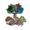
| Details | The electron density was clearly interpretable, which allowed us to build a de novo structural model. This process began by fitting the crystallographic model of the X. citri VirB7 C-terminal N0 domain (PDB:3OV5) and the NMR model of the X. citri VirB9CTD-VirB7NTD complex (PDB:2N01) in order to identify the map with the correct handedness. Models were positioned using Fit in map tool in Chimera, and saved relative to the map. Using these as starting points, we were able to manually build the rest of the model for VirB7 and VirB9CTD, and the de novo models for VirB10CTD, VirB10NTD_150-161 and VirB9NTD using Coot. In this manner, we obtained a combined model for a single VirB7-VirB9-VirB10 heterotrimer unit, which was submitted to iterative rounds of real space refinement and building using PHENIX and Coot software, respectively. Thirteen more copies of the refined heterotrimer were then fit into the density map using Chimera and new rounds of real space refinement (now using NCS for the 42 chains contained in the structure) and building using PHENIX and Coot, respectively, were executed until we obtained good parameters for Ramachandran plot and MolProbity. Chimera and PyMol were used for map and model visualization and figure production. |
|---|---|
| Refinement | Space: REAL / Overall B value: 138 / Target criteria: Cross-correlation coefficient |
| Output model | PDB-6gyb: |
