National Institutes of Health/National Institute of General Medical Sciences (NIH/NIGMS)
GM079179
米国
Welch Foundation
I-1578
米国
引用
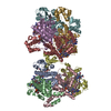
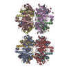
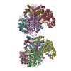
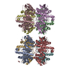
ジャーナル: Nature / 年: 2018 タイトル: Cryo-EM structure of a fungal mitochondrial calcium uniporter. 著者: Nam X Nguyen / Jean-Paul Armache / Changkeun Lee / Yi Yang / Weizhong Zeng / Vamsi K Mootha / Yifan Cheng / Xiao-Chen Bai / Youxing Jiang / 要旨: The mitochondrial calcium uniporter (MCU) is a highly selective calcium channel localized to the inner mitochondrial membrane. Here, we describe the structure of an MCU orthologue from the fungus ...The mitochondrial calcium uniporter (MCU) is a highly selective calcium channel localized to the inner mitochondrial membrane. Here, we describe the structure of an MCU orthologue from the fungus Neosartorya fischeri (NfMCU) determined to 3.8 Å resolution by phase-plate cryo-electron microscopy. The channel is a homotetramer with two-fold symmetry in its amino-terminal domain (NTD) that adopts a similar structure to that of human MCU. The NTD assembles as a dimer of dimers to form a tetrameric ring that connects to the transmembrane domain through an elongated coiled-coil domain. The ion-conducting pore domain maintains four-fold symmetry, with the selectivity filter positioned at the start of the pore-forming TM2 helix. The aspartate and glutamate sidechains of the conserved DIME motif are oriented towards the central axis and separated by one helical turn. The structure of NfMCU offers insights into channel assembly, selective calcium permeation, and inhibitor binding.
履歴
登録
2018年4月25日
登録サイト: RCSB / 処理サイト: RCSB
改定 1.0
2018年7月11日
Provider: repository / タイプ: Initial release
改定 1.1
2018年7月18日
Group: Data collection / Data processing / カテゴリ: em_3d_reconstruction / Item: _em_3d_reconstruction.resolution
根拠: Assembly of the saposin A molecules was determined based on clear electron density in the cryo-EM map, low pass-filtered to 5 Angstrom resolution using the crystal structure of saposin A (PDB ...根拠: Assembly of the saposin A molecules was determined based on clear electron density in the cryo-EM map, low pass-filtered to 5 Angstrom resolution using the crystal structure of saposin A (PDB ID: 2DOB) as the starting model for model building and refinement.
pH: 7.5 詳細: 20 mM HEPES, pH 7.5, 300 mM sodium chloride, 1 mM calcium chloride, 2% glycerol
試料
濃度: 0.6 mg/ml / 包埋: NO / シャドウイング: NO / 染色: NO / 凍結: YES 詳細: N. fischeri MCU was reconstituted into lipid using E. coli total lipids and human saposin A as the membrane scaffolding protein.
 ムービー
ムービー コントローラー


 データを開く
 基本情報
基本情報 要素
要素 キーワード
キーワード TRANSPORT PROTEIN (運搬体タンパク質) /
  機能・相同性情報

 データ登録者
 米国, 3件
  引用
引用 構造の表示
構造の表示 ダウンロードとリンク
 その他のダウンロード


 PDBj
PDBj 集合体
集合体


 分子量: 40.078 Da / 分子数: 1 / 由来タイプ: 合成 / 式: Ca
 試料調製
試料調製 電子顕微鏡撮影
 解析
解析