 Movie
Movie Controller

[English] 日本語
 Yorodumi
- EMDB-7828: Cryo-EM structure of the mitochondrial calcium uniporter from N. ... -
+ Open data

- Basic information
| Entry | Database: EMDB / ID: EMD-7828 | ||||||||||||
|---|---|---|---|---|---|---|---|---|---|---|---|---|---|
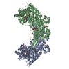
| Title | Cryo-EM structure of the mitochondrial calcium uniporter from N. fischeri bound to saposin | ||||||||||||
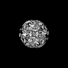
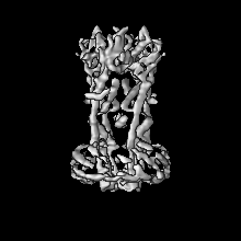
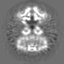
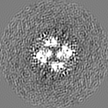
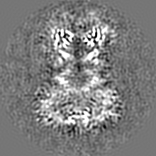
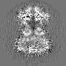
 Map data Map data | Cryo-EM structure of MCU from N. fischeri low pass-filtered to 5.0 Angstrom resolution to show saposin molecules | ||||||||||||
 Sample Sample |
| ||||||||||||
 Keywords Keywords | Mitochondria / calcium channel / TRANSPORT PROTEIN | ||||||||||||
| Function / homology |  Function and homology information Function and homology informationuniporter activity / uniplex complex / mitochondrial calcium ion homeostasis / calcium import into the mitochondrion / calcium channel activity / protein homotetramerization / mitochondrial inner membrane / metal ion binding Similarity search - Function | ||||||||||||
| Biological species |   Homo sapiens (human) Homo sapiens (human) | ||||||||||||
| Method | single particle reconstruction / cryo EM / Resolution: 5.0 Å | ||||||||||||
 Authors Authors | Nguyen NX / Armache J-P | ||||||||||||
| Funding support |  United States, 3 items United States, 3 items
| ||||||||||||
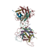
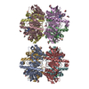
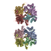
 Citation Citation | Journal: Nature / Year: 2018 Title: Cryo-EM structure of a fungal mitochondrial calcium uniporter. Authors: Nam X Nguyen / Jean-Paul Armache / Changkeun Lee / Yi Yang / Weizhong Zeng / Vamsi K Mootha / Yifan Cheng / Xiao-Chen Bai / Youxing Jiang / Abstract: The mitochondrial calcium uniporter (MCU) is a highly selective calcium channel localized to the inner mitochondrial membrane. Here, we describe the structure of an MCU orthologue from the fungus ...The mitochondrial calcium uniporter (MCU) is a highly selective calcium channel localized to the inner mitochondrial membrane. Here, we describe the structure of an MCU orthologue from the fungus Neosartorya fischeri (NfMCU) determined to 3.8 Å resolution by phase-plate cryo-electron microscopy. The channel is a homotetramer with two-fold symmetry in its amino-terminal domain (NTD) that adopts a similar structure to that of human MCU. The NTD assembles as a dimer of dimers to form a tetrameric ring that connects to the transmembrane domain through an elongated coiled-coil domain. The ion-conducting pore domain maintains four-fold symmetry, with the selectivity filter positioned at the start of the pore-forming TM2 helix. The aspartate and glutamate sidechains of the conserved DIME motif are oriented towards the central axis and separated by one helical turn. The structure of NfMCU offers insights into channel assembly, selective calcium permeation, and inhibitor binding. | ||||||||||||
| History |
|
- Structure visualization
| Movie |
Movie viewer |
|---|---|
| Structure viewer | EM map: SurfViewMolmilJmol/JSmol |
| Supplemental images |

- Downloads & links
-EMDB archive
| Map data | emd_7828.map.gz | 36.9 MB | EMDB map data format | |
|---|---|---|---|---|
| Header (meta data) | emd-7828-v30.xmlemd-7828.xml | 21.8 KB 21.8 KB | Display Display | EMDB header |
| Images | 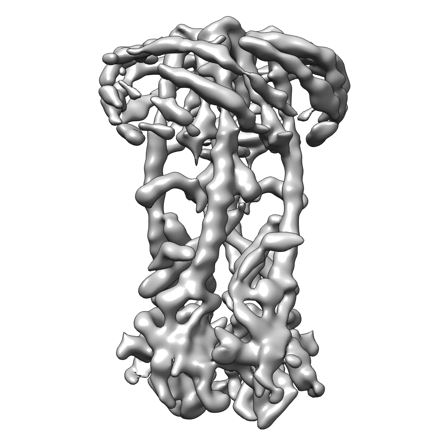 emd_7828.png emd_7828.png | 553.2 KB | ||
| Filedesc metadata | emd-7828.cif.gz | 6.8 KB | ||
| Others | emd_7828_additional.map.gzemd_7828_additional_1.map.gz | 37.2 MB 37.2 MB | ||
| Archive directory |  http://ftp.pdbj.org/pub/emdb/structures/EMD-7828ftp://ftp.pdbj.org/pub/emdb/structures/EMD-7828 http://ftp.pdbj.org/pub/emdb/structures/EMD-7828ftp://ftp.pdbj.org/pub/emdb/structures/EMD-7828 | HTTPS FTP |
-Related structure data
| Related structure data |  6d80MC  7826C  6d7wC C: citing same article ( M: atomic model generated by this map |
|---|---|
| Similar structure data |

-Links
| EMDB pages | EMDB (EBI/PDBe) / EMDataResource |
|---|
-Map
| File | Download / File: emd_7828.map.gz / Format: CCP4 / Size: 40.6 MB / Type: IMAGE STORED AS FLOATING POINT NUMBER (4 BYTES) | ||||||||||||||||||||||||||||||||||||||||||||||||||||||||||||||||||||
|---|---|---|---|---|---|---|---|---|---|---|---|---|---|---|---|---|---|---|---|---|---|---|---|---|---|---|---|---|---|---|---|---|---|---|---|---|---|---|---|---|---|---|---|---|---|---|---|---|---|---|---|---|---|---|---|---|---|---|---|---|---|---|---|---|---|---|---|---|---|
| Annotation | Cryo-EM structure of MCU from N. fischeri low pass-filtered to 5.0 Angstrom resolution to show saposin molecules | ||||||||||||||||||||||||||||||||||||||||||||||||||||||||||||||||||||
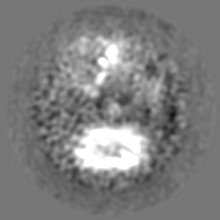
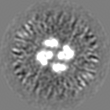
| Projections & slices | Image control
Images are generated by Spider. | ||||||||||||||||||||||||||||||||||||||||||||||||||||||||||||||||||||
| Voxel size | X=Y=Z: 0.84 Å | ||||||||||||||||||||||||||||||||||||||||||||||||||||||||||||||||||||
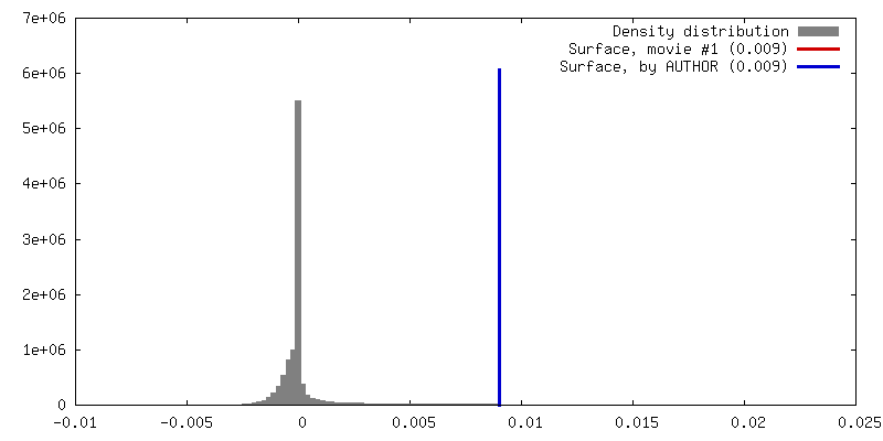
| Density |
| ||||||||||||||||||||||||||||||||||||||||||||||||||||||||||||||||||||
| Symmetry | Space group: 1 | ||||||||||||||||||||||||||||||||||||||||||||||||||||||||||||||||||||
| Details | EMDB XML:
CCP4 map header:
| ||||||||||||||||||||||||||||||||||||||||||||||||||||||||||||||||||||
 Z (Sec.)
 Y (Row.)
 X (Col.)


-Supplemental data
-Additional map: Cryo-EM structure of MCU from N. fischeri at 3.8 Angstrom resolution
| File | emd_7828_additional.map | ||||||||||||
|---|---|---|---|---|---|---|---|---|---|---|---|---|---|
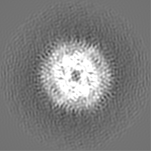
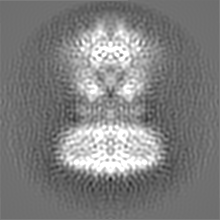
| Annotation | Cryo-EM structure of MCU from N. fischeri at 3.8 Angstrom resolution | ||||||||||||
| Projections & Slices |
| ||||||||||||
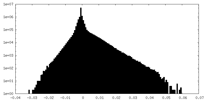
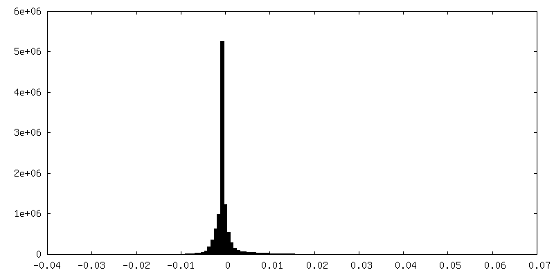
| Density Histograms |


-Additional map: Cryo-EM structure of MCU from N. fischeri at 3.8 Angstrom resolution
| File | emd_7828_additional_1.map | ||||||||||||
|---|---|---|---|---|---|---|---|---|---|---|---|---|---|
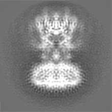
| Annotation | Cryo-EM structure of MCU from N. fischeri at 3.8 Angstrom resolution | ||||||||||||
| Projections & Slices |
| ||||||||||||
| Density Histograms |


- Sample components
-Entire : Mitochondrial calcium uniporter / saposin complex
| Entire | Name: Mitochondrial calcium uniporter / saposin complex |
|---|---|
| Components |
|
-Supramolecule #1: Mitochondrial calcium uniporter / saposin complex
| Supramolecule | Name: Mitochondrial calcium uniporter / saposin complex / type: organelle_or_cellular_component / ID: 1 / Parent: 0 / Macromolecule list: #1-#2 |
|---|
-Supramolecule #3: Mitochondrial calcium uniporter
| Supramolecule | Name: Mitochondrial calcium uniporter / type: organelle_or_cellular_component / ID: 3 / Parent: 1 / Macromolecule list: #2 |
|---|---|
| Source (natural) | Organism: |
-Supramolecule #2: saposin
| Supramolecule | Name: saposin / type: complex / ID: 2 / Parent: 1 / Macromolecule list: #1 |
|---|---|
| Source (natural) | Organism: Homo sapiens (human) |
-Macromolecule #1: Saposin A
| Macromolecule | Name: Saposin A / type: protein_or_peptide / ID: 1 / Number of copies: 6 / Enantiomer: LEVO |
|---|---|
| Source (natural) | Organism: Homo sapiens (human) |
| Molecular weight | Theoretical: 6.911511 KDa |
| Recombinant expression | Organism:  |
| Sequence | String: (UNK)(UNK)(UNK)(UNK)(UNK)(UNK)(UNK)(UNK)(UNK)(UNK) (UNK)(UNK)(UNK)(UNK)(UNK)(UNK) (UNK)(UNK)(UNK) (UNK)(UNK)(UNK)(UNK)(UNK)(UNK)(UNK)(UNK)(UNK)(UNK) (UNK)(UNK)(UNK) (UNK)(UNK)(UNK)(UNK)(UNK) ...String: (UNK)(UNK)(UNK)(UNK)(UNK)(UNK)(UNK)(UNK)(UNK)(UNK) (UNK)(UNK)(UNK)(UNK)(UNK)(UNK) (UNK)(UNK)(UNK) (UNK)(UNK)(UNK)(UNK)(UNK)(UNK)(UNK)(UNK)(UNK)(UNK) (UNK)(UNK)(UNK) (UNK)(UNK)(UNK)(UNK)(UNK)(UNK) (UNK)(UNK)(UNK)(UNK)(UNK)(UNK)(UNK)(UNK)(UNK)(UNK) (UNK)(UNK)(UNK)(UNK)(UNK)(UNK)(UNK)(UNK)(UNK) (UNK)(UNK)(UNK)(UNK)(UNK)(UNK)(UNK) (UNK)(UNK) (UNK)(UNK)(UNK)(UNK)(UNK)(UNK)(UNK)(UNK)(UNK)(UNK) (UNK)(UNK)(UNK)(UNK) (UNK) |
-Macromolecule #2: Mitochondrial calcium uniporter
| Macromolecule | Name: Mitochondrial calcium uniporter / type: protein_or_peptide / ID: 2 / Number of copies: 4 / Enantiomer: LEVO |
|---|---|
| Source (natural) | Organism: |
| Molecular weight | Theoretical: 47.928004 KDa |
| Recombinant expression | Organism: |
| Sequence | String: GSDKRGPQSA EPDPLERLEV KKVQQQHENE KDDSGRDTKS GGKVAKAMTK GDTIAGKLLT TPSRLFKLLI PLTTINRKDI EQIAILIHP QQPLSHLERL IQSEVPPIED ENGQKRPPFV SFIALQLEQD AIRPKRGMYE GTDAEIHRVE GGKDDATVAK R GEDFQEVD ...String: GSDKRGPQSA EPDPLERLEV KKVQQQHENE KDDSGRDTKS GGKVAKAMTK GDTIAGKLLT TPSRLFKLLI PLTTINRKDI EQIAILIHP QQPLSHLERL IQSEVPPIED ENGQKRPPFV SFIALQLEQD AIRPKRGMYE GTDAEIHRVE GGKDDATVAK R GEDFQEVD ETFSYLRRPG PGQGDKEQRF IRWSQSTEIG DFIRDAARAK EFIVTIEGAP AGLEQIHVAV PSFDERTYFL RM RLRKISR RIQGLAEIKH ECDALAHRGA QRVALGGFGI LAFWWYIVYK LTFETDLGWD TMEPVTYLVS LSTLMGGYLW FLY HNREIS YRSALDFTIN ARQKKLYQMK GIDLQVWESL IDEANAIRRE IKNIAAEYDV DWDERKDEQD DRVTEALKKE RRLK NGSQK EERPKDDRDD D UniProtKB: Calcium uniporter protein, mitochondrial |
-Macromolecule #3: CALCIUM ION
| Macromolecule | Name: CALCIUM ION / type: ligand / ID: 3 / Number of copies: 1 / Formula: CA |
|---|---|
| Molecular weight | Theoretical: 40.078 Da |
-Experimental details
-Structure determination
| Method | cryo EM |
|---|---|
 Processing Processing | single particle reconstruction |
| Aggregation state | particle |
-Sample preparation
| Concentration | 0.6 mg/mL |
|---|---|
| Buffer | pH: 7.5 Details: 20 mM HEPES, pH 7.5, 300 mM sodium chloride, 1 mM calcium chloride, 2% glycerol |
| Grid | Model: Quantifoil R1.2/1.3 / Material: GOLD / Mesh: 300 |
| Vitrification | Cryogen name: ETHANE / Chamber humidity: 100 % / Chamber temperature: 277 K / Instrument: FEI VITROBOT MARK IV |
| Details | N. fischeri MCU was reconstituted into lipid using E. coli total lipids and human saposin A as the membrane scaffolding protein. |
- Electron microscopy
| Microscope | FEI TITAN KRIOS |
|---|---|
| Specialist optics | Phase plate: VOLTA PHASE PLATE |
| Image recording | Film or detector model: GATAN K2 SUMMIT (4k x 4k) / Average electron dose: 60.0 e/Å2 |
| Electron beam | Acceleration voltage: 300 kV / Electron source:  FIELD EMISSION GUN FIELD EMISSION GUN |
| Electron optics | Illumination mode: FLOOD BEAM / Imaging mode: BRIGHT FIELD |
| Experimental equipment |  Model: Titan Krios / Image courtesy: FEI Company |
