| Entry | Database: PDB / ID: 6bsn
|
|---|
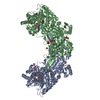
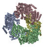
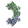
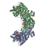
| Title | Structure of proline utilization A (PutA) with proline bound in remote sites |
|---|
 Components Components | Bifunctional protein PutA |
|---|
 Keywords Keywords | OXIDOREDUCTASE / FLAVOENZYME / ROSSMANN FOLD / ALDEHYDE DEHYDROGENASE / FLAVIN ADENINE DINUCLEOTIDE / NICOTINAMIDE ADENINE DINUCLEOTIDE / PROLINE CATABOLISM / SUBSTRATE CHANNELING / BIFUNCTIONAL ENZYME |
|---|
| Function / homology |  Function and homology information Function and homology information
proline dehydrogenase / proline dehydrogenase activity / L-glutamate gamma-semialdehyde dehydrogenase / L-glutamate gamma-semialdehyde dehydrogenase activity / : / cytoplasmic side of plasma membrane / DNA-binding transcription factor activity / nucleotide binding / DNA binding / identical protein bindingSimilarity search - Function TIM Barrel - #220 / Delta-1-pyrroline-5-carboxylate dehydrogenase 3 / Proline dehydrogenase PutA, domain II / Proline dehydrogenase PutA, domain I/II / DNA-binding domain of Proline dehydrogenase / Bifunctional protein PutA / Proline dehydrogenase domain / Proline dehydrogenase / : / FAD-linked oxidoreductase-like ...TIM Barrel - #220 / Delta-1-pyrroline-5-carboxylate dehydrogenase 3 / Proline dehydrogenase PutA, domain II / Proline dehydrogenase PutA, domain I/II / DNA-binding domain of Proline dehydrogenase / Bifunctional protein PutA / Proline dehydrogenase domain / Proline dehydrogenase / : / FAD-linked oxidoreductase-like / Aldehyde Dehydrogenase; Chain A, domain 2 / Aldehyde Dehydrogenase; Chain A, domain 2 / Aldehyde Dehydrogenase; Chain A, domain 1 / Aldehyde Dehydrogenase; Chain A, domain 1 / Aldehyde dehydrogenase, cysteine active site / Aldehyde dehydrogenases cysteine active site. / Aldehyde dehydrogenase domain / Aldehyde dehydrogenase family / Aldehyde dehydrogenase, C-terminal / Aldehyde dehydrogenase, N-terminal / Aldehyde/histidinol dehydrogenase / TIM Barrel / Alpha-Beta Barrel / 3-Layer(aba) Sandwich / Alpha BetaSimilarity search - Domain/homology |
|---|
| Biological species |  Bradyrhizobium diazoefficiens (bacteria) Bradyrhizobium diazoefficiens (bacteria) |
|---|
| Method |  X-RAY DIFFRACTION / SYNCHROTRON / FOURIER SYNTHESIS / Resolution: 2.15 Å X-RAY DIFFRACTION / SYNCHROTRON / FOURIER SYNTHESIS / Resolution: 2.15 Å |
|---|
 Authors Authors | Tanner, J.J. / Korasick, D.A. |
|---|
| Funding support |  United States, 2items United States, 2items | Organization | Grant number | Country |
|---|
| National Institutes of Health/National Institute of General Medical Sciences (NIH/NIGMS) | R01GM065546 | United States | | National Institutes of Health/National Institute of General Medical Sciences (NIH/NIGMS) | R01GM061068 | United States |
|
|---|
 Citation Citation | Journal: Molecules / Year: 2017
Title: Structural Basis for the Substrate Inhibition of Proline Utilization A by Proline.
Authors: Korasick, D.A. / Pemberton, T.A. / Arentson, B.W. / Becker, D.F. / Tanner, J.J. |
|---|
| History | | Deposition | Dec 4, 2017 | Deposition site: RCSB / Processing site: RCSB |
|---|
| Revision 1.0 | Jan 3, 2018 | Provider: repository / Type: Initial release |
|---|
| Revision 1.1 | Jan 31, 2018 | Group: Database references / Category: citation
Item: _citation.country / _citation.journal_abbrev ..._citation.country / _citation.journal_abbrev / _citation.journal_id_ASTM / _citation.journal_id_CSD / _citation.journal_id_ISSN / _citation.page_first / _citation.pdbx_database_id_PubMed / _citation.title / _citation.year |
|---|
| Revision 1.2 | Jan 1, 2020 | Group: Author supporting evidence / Category: pdbx_audit_support / Item: _pdbx_audit_support.funding_organization |
|---|
| Revision 1.3 | Oct 4, 2023 | Group: Data collection / Database references / Refinement description
Category: chem_comp_atom / chem_comp_bond ...chem_comp_atom / chem_comp_bond / database_2 / pdbx_initial_refinement_model
Item: _database_2.pdbx_DOI / _database_2.pdbx_database_accession |
|---|
|
|---|
 Movie
Movie Controller

 Yorodumi
 Open data
 Basic information
 Structure visualization
 Downloads & links
 Other downloads


 PDBj
PDBj


 Assembly