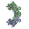
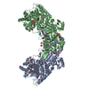
Prolinedehydrogenase / Proline utilization A (PutA) bifunctional proline dehydrogenase and 1-pyrroline-5-carboxylate dehydrogenase
Mass: 107697.844 Da / Num. of mol.: 2 Source method: isolated from a genetically manipulated source Details: Gly-His at the N-terminus is a cloning artifact. Source: (gene. exp.) Bradyrhizobium japonicum USDA 110 (bacteria) Gene: blr7261, putA / Plasmid: pKA8H / Production host: Escherichia coli (E. coli) / References: UniProt: Q89E26, EC: 1.5.99.8, EC: 1.5.1.12
Mass: 18.015 Da / Num. of mol.: 795 / Source method: isolated from a natural source / Formula: H2O
-
Experimental details
-
Experiment
Experiment
Method: X-RAY DIFFRACTION / Number of used crystals: 1
-
Sample preparation
Crystal
Density Matthews: 3.51 Å3/Da / Density % sol: 64.99 %
Crystal grow
Temperature: 298 K / Method: vapor diffusion, sitting drop / pH: 7 Details: 2M Ammonium sulfate, 0.1 M Tris-HCl pH 7.0. N-terminal His tag cleaved by TEV protease, which leaves Gly-His at N-terminus., VAPOR DIFFUSION, SITTING DROP, temperature 298K
-
Data collection
Diffraction
Mean temperature: 100 K
Diffraction source
Source: SYNCHROTRON / Site: ALS / Beamline: 4.2.2 / Wavelength: 1 Å
Detector
Type: NOIR-1 / Detector: CCD / Date: May 2, 2007
Radiation
Monochromator: beamline optics / Protocol: SINGLE WAVELENGTH / Monochromatic (M) / Laue (L): M / Scattering type: x-ray
Radiation wavelength
Wavelength: 1 Å / Relative weight: 1
Reflection
Resolution: 2.1→43.27 Å / Num. obs: 172549 / % possible obs: 100 % / Redundancy: 3.8 % / Rmerge(I) obs: 0.103 / Rsym value: 0.103 / Net I/σ(I): 5.386
Reflection shell
Resolution (Å)
Redundancy (%)
Rmerge(I) obs
Mean I/σ(I) obs
Num. measured all
Num. unique all
Rsym value
% possible all
2.1-2.21
3.7
0.412
1.8
93890
25188
0.412
100
2.21-2.35
3.8
0.313
2.1
89339
23780
0.313
100
2.35-2.51
3.8
0.239
3.1
84702
22410
0.239
100
2.51-2.71
3.8
0.182
4.1
79097
20815
0.182
100
2.71-2.97
3.8
0.131
5.6
73120
19140
0.131
100
2.97-3.32
3.8
0.096
7.4
66528
17371
0.096
100
3.32-3.83
3.8
0.072
8.9
58632
15304
0.072
100
3.83-4.7
3.8
0.065
9.2
49733
12965
0.065
100
4.7-6.64
3.8
0.064
9.4
38421
10030
0.064
100
6.64-43.27
3.7
0.07
7.2
20641
5546
0.07
99.6
-
Processing
Software
Name
Version
Classification
NB
SCALA
3.2.25
dataprocessing
PHENIX
refinement
PDB_EXTRACT
3.005
dataextraction
MOSFLM
datareduction
SCALA
datascaling
SOLVE
phasing
RESOLVE
phasing
MOLREP
phasing
Refinement
Method to determine structure: combination of Se-Met MAD/SAD, molecular replacement Starting model: Partial model from MAD/SAD phasing from another crystal form Resolution: 2.1→42.33 Å / Occupancy max: 1 / Occupancy min: 0.45 / FOM work R set: 0.851 / SU ML: 0.67 / Stereochemistry target values: MAXIMUM LIKELIHOOD
Rfactor
Num. reflection
% reflection
Selection details
Rfree
0.233
8703
5.04 %
random
Rwork
0.2
-
-
-
obs
0.202
172521
99.98 %
-
Solvent computation
Shrinkage radii: 0.9 Å / VDW probe radii: 1.11 Å / Solvent model: FLAT BULK SOLVENT MODEL / Bsol: 50.55 Å2 / ksol: 0.383 e/Å3
In the structure databanks used in Yorodumi, some data are registered as the other names, "COVID-19 virus" and "2019-nCoV". Here are the details of the virus and the list of structure data.
Jan 31, 2019. EMDB accession codes are about to change! (news from PDBe EMDB page)
EMDB accession codes are about to change! (news from PDBe EMDB page)
The allocation of 4 digits for EMDB accession codes will soon come to an end. Whilst these codes will remain in use, new EMDB accession codes will include an additional digit and will expand incrementally as the available range of codes is exhausted. The current 4-digit format prefixed with “EMD-” (i.e. EMD-XXXX) will advance to a 5-digit format (i.e. EMD-XXXXX), and so on. It is currently estimated that the 4-digit codes will be depleted around Spring 2019, at which point the 5-digit format will come into force.
The EM Navigator/Yorodumi systems omit the EMD- prefix.
Related info.:Q: What is EMD? / ID/Accession-code notation in Yorodumi/EM Navigator
Yorodumi is a browser for structure data from EMDB, PDB, SASBDB, etc.
This page is also the successor to EM Navigator detail page, and also detail information page/front-end page for Omokage search.
The word "yorodu" (or yorozu) is an old Japanese word meaning "ten thousand". "mi" (miru) is to see.
Related info.:EMDB / PDB / SASBDB / Comparison of 3 databanks / Yorodumi Search / Aug 31, 2016. New EM Navigator & Yorodumi / Yorodumi Papers / Jmol/JSmol / Function and homology information / Changes in new EM Navigator and Yorodumi
 Movie
Movie Controller

 Yorodumi
 Open data
 Basic information
 Components
 Keywords
 Function and homology information
 Bradyrhizobium japonicum USDA 110 (bacteria)
 X-RAY DIFFRACTION /
  Authors
 Citation
 Structure visualization
 Downloads & links
 Other downloads
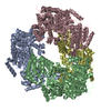


 PDBj
PDBj

 Assembly


 Mass: 785.550 Da / Num. of mol.: 2 / Source method: obtained synthetically / Formula: C27H33N9O15P2 / Comment: FAD*YM
 Mass: 663.425 Da / Num. of mol.: 2 / Source method: obtained synthetically / Formula: C21H27N7O14P2 / Comment: NAD*YM
 Mass: 96.063 Da / Num. of mol.: 13 / Source method: obtained synthetically / Formula: SO4
 Mass: 92.094 Da / Num. of mol.: 6 / Source method: obtained synthetically / Formula: C3H8O3
 Sample preparation
 / Beamline: 4.2.2 / Wavelength: 1 Å
 Processing