 ムービー
ムービー コントローラー


+ データを開く

- 基本情報
基本情報
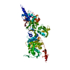
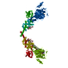
| 登録情報 | データベース: PDB / ID: 5hlb | ||||||||||||||||||
|---|---|---|---|---|---|---|---|---|---|---|---|---|---|---|---|---|---|---|---|
| タイトル | E. coli PBP1b in complex with acyl-aztreonam and moenomycin | ||||||||||||||||||
 要素 要素 | Penicillin-binding protein 1B | ||||||||||||||||||
 キーワード キーワード | TRANSFERASE/TRANSFERASE INHIBITOR /  penicillin-binding protein (ペニシリン結合タンパク質) / inhibitor complex (酵素阻害剤) / transpeptidase / transglycosylase (グリコシルトランスフェラーゼ) / TRANSFERASE-TRANSFERASE INHIBITOR complex penicillin-binding protein (ペニシリン結合タンパク質) / inhibitor complex (酵素阻害剤) / transpeptidase / transglycosylase (グリコシルトランスフェラーゼ) / TRANSFERASE-TRANSFERASE INHIBITOR complex | ||||||||||||||||||
| 機能・相同性 |  機能・相同性情報 機能・相同性情報positive regulation of bipolar cell growth / cell wall repair / peptidoglycan glycosyltransferase / peptidoglycan glycosyltransferase activity / serine-type D-Ala-D-Ala carboxypeptidase / serine-type D-Ala-D-Ala carboxypeptidase activity / penicillin binding / peptidoglycan biosynthetic process / peptidoglycan-based cell wall / outer membrane-bounded periplasmic space ...positive regulation of bipolar cell growth / cell wall repair / peptidoglycan glycosyltransferase / peptidoglycan glycosyltransferase activity / serine-type D-Ala-D-Ala carboxypeptidase / serine-type D-Ala-D-Ala carboxypeptidase activity / penicillin binding / peptidoglycan biosynthetic process / peptidoglycan-based cell wall / outer membrane-bounded periplasmic space / regulation of cell shape / response to antibiotic / タンパク質分解 / 生体膜 / 細胞膜類似検索 - 分子機能 | ||||||||||||||||||
| 生物種 |  Escherichia coli (大腸菌) Escherichia coli (大腸菌) | ||||||||||||||||||
| 手法 | X線回折 / シンクロトロン / 分子置換 / 解像度: 2.42 Å | ||||||||||||||||||
 データ登録者 データ登録者 | King, D.T. / Strynadka, N.C.J. | ||||||||||||||||||
| 資金援助 |  カナダ, カナダ,  米国, 5件 米国, 5件
| ||||||||||||||||||
 引用 引用 | ジャーナル: J. Biol. Chem. / 年: 2016 タイトル: Escherichia coli Penicillin-Binding Protein 1B: Structural Insights into Inhibition. 著者: King, D.T. / Wasney, G.A. / Nosella, M. / Fong, A. / Strynadka, N.C. | ||||||||||||||||||
| 履歴 |
|
- 構造の表示
構造の表示
| 構造ビューア | 分子: MolmilJmol/JSmol |
|---|
- ダウンロードとリンク
-ダウンロード
| PDBx/mmCIF形式 | 5hlb.cif.gz | 171.2 KB | 表示 | PDBx/mmCIF形式 |
|---|---|---|---|---|
| PDB形式 | pdb5hlb.ent.gz | 129.5 KB | 表示 | PDB形式 |
| PDBx/mmJSON形式 | 5hlb.json.gz | ツリー表示 | PDBx/mmJSON形式 | |
| その他 |  その他のダウンロード その他のダウンロード |
-検証レポート
| アーカイブディレクトリ | https://data.pdbj.org/pub/pdb/validation_reports/hl/5hlbftp://data.pdbj.org/pub/pdb/validation_reports/hl/5hlb | HTTPS FTP |
|---|
-関連構造データ


-リンク
 PDBj
PDBj
- 集合体
集合体
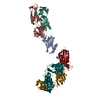
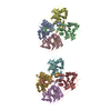
| 登録構造単位 | 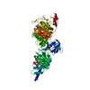
| ||||||||
|---|---|---|---|---|---|---|---|---|---|
| 1 | 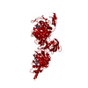
| ||||||||
| 単位格子 |
|
-要素
| #1: タンパク質 | 分子量: 83280.352 Da / 分子数: 1 / 由来タイプ: 組換発現 由来: (組換発現) Escherichia coli (strain K12) (大腸菌)株: K12 / 遺伝子: mrcB, pbpF, ponB, b0149, JW0145 / 発現宿主: Escherichia coli (大腸菌)参照: UniProt: P02919, peptidoglycan glycosyltransferase, 加水分解酵素; プロテアーゼ; ペプチド結合加水分解酵素 |
|---|---|
| #2: 化合物 | ChemComp-M0E / Moenomycin family antibiotics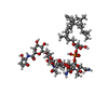  分子量: 1580.567 Da / 分子数: 1 / 由来タイプ: 合成 / 式: C69H106N5O34P / コメント: 抗生剤*YM 分子量: 1580.567 Da / 分子数: 1 / 由来タイプ: 合成 / 式: C69H106N5O34P / コメント: 抗生剤*YM |
| #3: 化合物 | ChemComp-AZR / アズトレオナム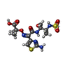  分子量: 437.449 Da / 分子数: 1 / 由来タイプ: 合成 / 式: C13H19N5O8S2 / コメント: 抗生剤*YM 分子量: 437.449 Da / 分子数: 1 / 由来タイプ: 合成 / 式: C13H19N5O8S2 / コメント: 抗生剤*YM |
| #4: 水 | ChemComp-HOH / 水 分子量: 18.015 Da / 分子数: 233 / 由来タイプ: 天然 / 式: H2O 分子量: 18.015 Da / 分子数: 233 / 由来タイプ: 天然 / 式: H2O |
-実験情報
-実験
| 実験 | 手法: X線回折 / 使用した結晶の数: 1 |
|---|
- 試料調製
試料調製
| 結晶 | マシュー密度: 3.64 Å3/Da / 溶媒含有率: 66.24 % |
|---|---|
| 結晶化 | 温度: 298 K / 手法: 蒸気拡散法, シッティングドロップ法 詳細: Drops contained 1uL of protein solution (20mg/mL protein, 100uM moenomycin and 2mM aztreonam) mixed with an equal volume of precipitant (20% w/v PEG 3350, 0.2M potassium/sodium tartate, 0.1M Bis Tris pH 8.5). |
-データ収集
| 回折 | 平均測定温度: 100 K |
|---|---|
| 放射光源 | 由来: シンクロトロン / サイト: CLSI / ビームライン: 08ID-1 / 波長: 1 Å |
| 検出器 | タイプ: RAYONIX MX300HE / 検出器: CCD / 日付: 2013年2月23日 |
| 放射 | プロトコル: SINGLE WAVELENGTH / 単色(M)・ラウエ(L): M / 散乱光タイプ: x-ray |
| 放射波長 | 波長: 1 Å / 相対比: 1 |
| 反射 | 解像度: 2.42→64.39 Å / Num. obs: 45993 / % possible obs: 96.7 % / 冗長度: 3.5 % / Biso Wilson estimate: 44.38 Å2 / Rmerge(I) obs: 0.058 / Net I/σ(I): 12.7 |
| 反射 シェル | 解像度: 2.42→2.48 Å / Rmerge(I) obs: 0.552 |
- 解析
解析
| ソフトウェア |
| ||||||||||||||||||||||||||||||||||||||||||||||||||||||||||||||||||||||||||||||||||||||||||||||||||||||||||||||||||
|---|---|---|---|---|---|---|---|---|---|---|---|---|---|---|---|---|---|---|---|---|---|---|---|---|---|---|---|---|---|---|---|---|---|---|---|---|---|---|---|---|---|---|---|---|---|---|---|---|---|---|---|---|---|---|---|---|---|---|---|---|---|---|---|---|---|---|---|---|---|---|---|---|---|---|---|---|---|---|---|---|---|---|---|---|---|---|---|---|---|---|---|---|---|---|---|---|---|---|---|---|---|---|---|---|---|---|---|---|---|---|---|---|---|---|---|
| 精密化 | 構造決定の手法: 分子置換 開始モデル: 3VMA 解像度: 2.42→35.43 Å / Cor.coef. Fo:Fc: 0.8579 / Cor.coef. Fo:Fc free: 0.8491 / SU R Cruickshank DPI: 0.292 / 交差検証法: THROUGHOUT / σ(F): 0 / SU R Blow DPI: 0.298 / SU Rfree Blow DPI: 0.216 / SU Rfree Cruickshank DPI: 0.216
| ||||||||||||||||||||||||||||||||||||||||||||||||||||||||||||||||||||||||||||||||||||||||||||||||||||||||||||||||||
| 原子変位パラメータ | Biso mean: 61.29 Å2
| ||||||||||||||||||||||||||||||||||||||||||||||||||||||||||||||||||||||||||||||||||||||||||||||||||||||||||||||||||
| Refine analyze | Luzzati coordinate error obs: 0.374 Å | ||||||||||||||||||||||||||||||||||||||||||||||||||||||||||||||||||||||||||||||||||||||||||||||||||||||||||||||||||
| 精密化ステップ | サイクル: 1 / 解像度: 2.42→35.43 Å
| ||||||||||||||||||||||||||||||||||||||||||||||||||||||||||||||||||||||||||||||||||||||||||||||||||||||||||||||||||
| 拘束条件 |
| ||||||||||||||||||||||||||||||||||||||||||||||||||||||||||||||||||||||||||||||||||||||||||||||||||||||||||||||||||
| LS精密化 シェル | 解像度: 2.42→2.48 Å / Total num. of bins used: 20
|
