 Movie
Movie Controller


+ Open data

- Basic information
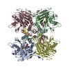
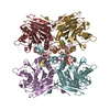
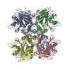
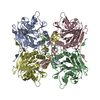
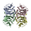
| Entry | Database: PDB / ID: 3pva | ||||||
|---|---|---|---|---|---|---|---|
| Title | PENICILLIN V ACYLASE FROM B. SPHAERICUS | ||||||
 Components Components | PROTEIN (PENICILLIN V ACYLASE) | ||||||
 Keywords Keywords | HYDROLASE / AMIDOHYDROLASE / NTN HYDROLASE / PENICILLIN V ACYLASE | ||||||
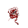
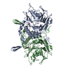
| Function / homology |  Function and homology information Function and homology informationpenicillin amidase activity / penicillin amidase / response to antibiotic Similarity search - Function | ||||||
| Biological species |  Lysinibacillus sphaericus (bacteria) Lysinibacillus sphaericus (bacteria) | ||||||
| Method |  X-RAY DIFFRACTION / MOLECULAR REPLACEMENT / Resolution: 2.8 Å X-RAY DIFFRACTION / MOLECULAR REPLACEMENT / Resolution: 2.8 Å | ||||||
 Authors Authors | Suresh, C.G. / Pundle, A.V. / Rao, K.N. / Sivaraman, H. / Brannigan, J.A. / Mcvey, C.E. / Verma, C.S. / Dauter, Z. / Dodson, E.J. / Dodson, G.G. | ||||||
 Citation Citation | Journal: Nat.Struct.Biol. / Year: 1999 Title: Penicillin V acylase crystal structure reveals new Ntn-hydrolase family members. Authors: Suresh, C.G. / Pundle, A.V. / SivaRaman, H. / Rao, K.N. / Brannigan, J.A. / McVey, C.E. / Verma, C.S. / Dauter, Z. / Dodson, E.J. / Dodson, G.G. | ||||||
| History |
|
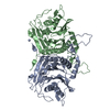
- Structure visualization
| Structure viewer | Molecule: MolmilJmol/JSmol |
|---|
- Downloads & links
-Download
| PDBx/mmCIF format | 3pva.cif.gz | 573.4 KB | Display | PDBx/mmCIF format |
|---|---|---|---|---|
| PDB format | pdb3pva.ent.gz | 470.9 KB | Display | PDB format |
| PDBx/mmJSON format | 3pva.json.gz | Tree view | PDBx/mmJSON format | |
| Others |  Other downloads Other downloads |
-Validation report
| Arichive directory | https://data.pdbj.org/pub/pdb/validation_reports/pv/3pvaftp://data.pdbj.org/pub/pdb/validation_reports/pv/3pva | HTTPS FTP |
|---|
-Related structure data

-Links
 PDBj
PDBj


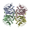
- Assembly
| Deposited unit | 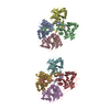
| ||||||||
|---|---|---|---|---|---|---|---|---|---|
| 1 | 
| ||||||||
| 2 | 
| ||||||||
| Unit cell |
|
-Components
| #1: Protein | Mass: 37189.105 Da / Num. of mol.: 8 / Source method: isolated from a natural source / Source: (natural) Lysinibacillus sphaericus (bacteria) / References: UniProt: P12256, penicillin amidase#2: Water | ChemComp-HOH / |  Mass: 18.015 Da / Num. of mol.: 2800 / Source method: isolated from a natural source / Formula: H2O Mass: 18.015 Da / Num. of mol.: 2800 / Source method: isolated from a natural source / Formula: H2O |
|---|
-Experimental details
-Experiment
| Experiment | Method: X-RAY DIFFRACTION / Number of used crystals: 1 |
|---|
- Sample preparation
| Crystal | Density Matthews: 3.2 Å3/Da / Density % sol: 61.54 % | ||||||||||||||||||||||||||||||||||||||||||
|---|---|---|---|---|---|---|---|---|---|---|---|---|---|---|---|---|---|---|---|---|---|---|---|---|---|---|---|---|---|---|---|---|---|---|---|---|---|---|---|---|---|---|---|
| Crystal grow | pH: 6.4 Details: 5MM DTT, 30% AS, 1% SUCROSE, 0.2M NA PHOSPHATE, PH 6.4 | ||||||||||||||||||||||||||||||||||||||||||
| Crystal grow | *PLUS Method: vapor diffusion, hanging drop | ||||||||||||||||||||||||||||||||||||||||||
| Components of the solutions | *PLUS
|
-Data collection
| Diffraction | Mean temperature: 120 K |
|---|---|
| Diffraction source | Source: ROTATING ANODE / Type: RIGAKU / Wavelength: 1.54 |
| Radiation | Protocol: SINGLE WAVELENGTH / Monochromatic (M) / Laue (L): M / Scattering type: x-ray |
| Radiation wavelength | Wavelength: 1.54 Å / Relative weight: 1 |
| Reflection | Resolution: 2.8→18 Å / Num. obs: 77056 / % possible obs: 85 % / Observed criterion σ(I): 0 / Redundancy: 2 % / Rmerge(I) obs: 0.053 / Net I/σ(I): 11.2 |
| Reflection | *PLUS Num. measured all: 111412 |
- Processing
| Software |
| |||||||||||||||||||||||||||||||||||||||||||||||||||||||||||||||
|---|---|---|---|---|---|---|---|---|---|---|---|---|---|---|---|---|---|---|---|---|---|---|---|---|---|---|---|---|---|---|---|---|---|---|---|---|---|---|---|---|---|---|---|---|---|---|---|---|---|---|---|---|---|---|---|---|---|---|---|---|---|---|---|---|
| Refinement | Method to determine structure: MOLECULAR REPLACEMENT Starting model: REFINED STRUCTURE FROM HEXAGONAL FORM Resolution: 2.8→14 Å / Cross valid method: THROUGHOUT / σ(F): 0
| |||||||||||||||||||||||||||||||||||||||||||||||||||||||||||||||
| Displacement parameters | Biso mean: 56.6 Å2 | |||||||||||||||||||||||||||||||||||||||||||||||||||||||||||||||
| Refinement step | Cycle: LAST / Resolution: 2.8→14 Å
| |||||||||||||||||||||||||||||||||||||||||||||||||||||||||||||||
| Refine LS restraints |
| |||||||||||||||||||||||||||||||||||||||||||||||||||||||||||||||
| Software | *PLUS Name: REFMAC / Classification: refinement | |||||||||||||||||||||||||||||||||||||||||||||||||||||||||||||||
| Refinement | *PLUS Highest resolution: 2.8 Å / Lowest resolution: 14 Å / σ(F): 0 / % reflection Rfree: 5 % / Rfactor obs: 0.211 | |||||||||||||||||||||||||||||||||||||||||||||||||||||||||||||||
| Solvent computation | *PLUS | |||||||||||||||||||||||||||||||||||||||||||||||||||||||||||||||
| Displacement parameters | *PLUS Biso mean: 56.6 Å2 | |||||||||||||||||||||||||||||||||||||||||||||||||||||||||||||||
| Refine LS restraints | *PLUS
|
