 Movie
Movie Controller

[English] 日本語
 Yorodumi
- PDB-2z71: Structure of truncated mutant CYS1GLY of penicillin V acylase fro... -
+ Open data

- Basic information
| Entry | Database: PDB / ID: 2z71 | ||||||
|---|---|---|---|---|---|---|---|
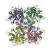
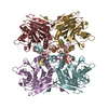
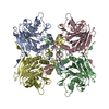
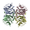
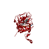
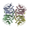
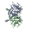
| Title | Structure of truncated mutant CYS1GLY of penicillin V acylase from bacillus sphaericus co-crystallized with penicillin V | ||||||
 Components Components | Penicillin acylase | ||||||
 Keywords Keywords | HYDROLASE / ZYMOGEN / PRECURSOR / PENICILLIN / AUTOPROTEOLYSIS / ANTIBIOTIC RESISTANCE / CATALYSIS / PENICILLIN V ACYLASE | ||||||
| Function / homology |  Function and homology information Function and homology informationpenicillin amidase activity / penicillin amidase / response to antibiotic Similarity search - Function | ||||||
| Biological species |  Bacillus sphaericus (bacteria) Bacillus sphaericus (bacteria) | ||||||
| Method |  X-RAY DIFFRACTION / SYNCHROTRON / MOLECULAR REPLACEMENT / Resolution: 2.6 Å X-RAY DIFFRACTION / SYNCHROTRON / MOLECULAR REPLACEMENT / Resolution: 2.6 Å | ||||||
 Authors Authors | Pathak, M.C. / Brannigan, J. / Dodson, G.G. / Suresh, C.G. | ||||||
 Citation Citation | Journal: To be published Title: Studies on the catalysis and post translational processing of penicillin V acylase Authors: Pathak, M.C. / Brannigan, J. / Dodson, G.G. / Suresh, C.G. #1: Journal: To be PublishedTitle: Auto-proteolytic processing of Penicillin V Acylase is simpler than of other related Ntn hydrolases Authors: Pathak, M.C. / Brannigan, J. / Dodson, G.G. / Suresh, C.G. #2: Journal: To be PublishedTitle: Co-crystal structure of Penicillin V Acylase with substrate Penicillin V: Insight in to catalytic specificity Authors: Pathak, M.C. / Brannigan, J. / Dodson, G.G. / Suresh, C.G. #3: Journal: To be PublishedTitle: Polymorphism shown by the crystals of Penicillin V Acyalse from Bacillus sphaericus Authors: Pathak, M.C. / Brannigan, J. / Dodson, G.G. / Suresh, C.G. | ||||||
| History |
|
- Structure visualization
| Structure viewer | Molecule: MolmilJmol/JSmol |
|---|
- Downloads & links
-Download
| PDBx/mmCIF format | 2z71.cif.gz | 140.4 KB | Display | PDBx/mmCIF format |
|---|---|---|---|---|
| PDB format | pdb2z71.ent.gz | 111.1 KB | Display | PDB format |
| PDBx/mmJSON format | 2z71.json.gz | Tree view | PDBx/mmJSON format | |
| Others |  Other downloads Other downloads |
-Validation report
| Arichive directory | https://data.pdbj.org/pub/pdb/validation_reports/z7/2z71ftp://data.pdbj.org/pub/pdb/validation_reports/z7/2z71 | HTTPS FTP |
|---|
-Related structure data
| Related structure data | 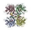 2pvaS S: Starting model for refinement |
|---|---|
| Similar structure data |


-Links
 PDBj
PDBj


- Assembly
| Deposited unit | 
| ||||||||
|---|---|---|---|---|---|---|---|---|---|
| 1 | 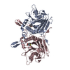
| ||||||||
| Unit cell |
|
-Components
| #1: Protein | Mass: 37199.105 Da / Num. of mol.: 2 / Mutation: C1G Source method: isolated from a genetically manipulated source Source: (gene. exp.) Bacillus sphaericus (bacteria) / Strain: NCIM 2478 / Production host: #2: Chemical | 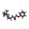  Mass: 350.390 Da / Num. of mol.: 2 / Source method: obtained synthetically / Formula: C16H18N2O5S / Comment: antibiotic*YM Mass: 350.390 Da / Num. of mol.: 2 / Source method: obtained synthetically / Formula: C16H18N2O5S / Comment: antibiotic*YM#3: Water | ChemComp-HOH / |  Mass: 18.015 Da / Num. of mol.: 51 / Source method: isolated from a natural source / Formula: H2O Mass: 18.015 Da / Num. of mol.: 51 / Source method: isolated from a natural source / Formula: H2O |
|---|
-Experimental details
-Experiment
| Experiment | Method: X-RAY DIFFRACTION / Number of used crystals: 1 |
|---|
- Sample preparation
| Crystal | Density Matthews: 3.14 Å3/Da / Density % sol: 60.86 % |
|---|---|
| Crystal grow | Method: vapor diffusion, hanging drop / pH: 6.4 / Details: pH 6.4, VAPOR DIFFUSION, HANGING DROP |
-Data collection
| Diffraction | Mean temperature: 100 K |
|---|---|
| Diffraction source | Source: SYNCHROTRON / Site: ESRF  / Beamline: ID14-1 / Wavelength: 0.933 Å / Beamline: ID14-1 / Wavelength: 0.933 Å |
| Detector | Date: Mar 8, 2003 |
| Radiation | Protocol: SINGLE WAVELENGTH / Monochromatic (M) / Laue (L): M / Scattering type: x-ray |
| Radiation wavelength | Wavelength: 0.933 Å / Relative weight: 1 |
| Reflection | Resolution: 2.6→20 Å / Num. all: 78639 / Num. obs: 28157 / % possible obs: 89.6 % |
- Processing
| Software |
| ||||||||||||||||||||||||||||||||||||||||||||||||||||||||||||||||||||||
|---|---|---|---|---|---|---|---|---|---|---|---|---|---|---|---|---|---|---|---|---|---|---|---|---|---|---|---|---|---|---|---|---|---|---|---|---|---|---|---|---|---|---|---|---|---|---|---|---|---|---|---|---|---|---|---|---|---|---|---|---|---|---|---|---|---|---|---|---|---|---|---|
| Refinement | Method to determine structure: MOLECULAR REPLACEMENT Starting model: 2PVA Resolution: 2.6→20 Å / Cor.coef. Fo:Fc: 0.936 / Cor.coef. Fo:Fc free: 0.914 / SU B: 13.545 / SU ML: 0.28 / Cross valid method: THROUGHOUT / ESU R: 0.743 / ESU R Free: 0.339 / Stereochemistry target values: MAXIMUM LIKELIHOOD
| ||||||||||||||||||||||||||||||||||||||||||||||||||||||||||||||||||||||
| Solvent computation | Ion probe radii: 0.8 Å / Shrinkage radii: 0.8 Å / VDW probe radii: 1.4 Å / Solvent model: BABINET MODEL WITH MASK | ||||||||||||||||||||||||||||||||||||||||||||||||||||||||||||||||||||||
| Displacement parameters | Biso mean: 46.447 Å2
| ||||||||||||||||||||||||||||||||||||||||||||||||||||||||||||||||||||||
| Refinement step | Cycle: LAST / Resolution: 2.6→20 Å
| ||||||||||||||||||||||||||||||||||||||||||||||||||||||||||||||||||||||
| Refine LS restraints |
| ||||||||||||||||||||||||||||||||||||||||||||||||||||||||||||||||||||||
| LS refinement shell | Resolution: 2.6→2.666 Å / Total num. of bins used: 20 /
|
