 ムービー
ムービー コントローラー


+ データを開く

- 基本情報
基本情報
| 登録情報 | データベース: PDB / ID: 4iim | ||||||
|---|---|---|---|---|---|---|---|
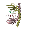
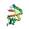
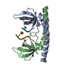
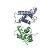
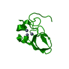
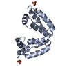
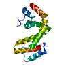
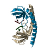
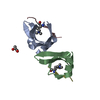
| タイトル | Crystal structure of the Second SH3 Domain of ITSN1 bound with a synthetic peptide | ||||||
 要素 要素 |
| ||||||
 キーワード キーワード |  ENDOCYTOSIS (エンドサイトーシス) / SH3 Domain (SH3ドメイン) / ITSN1 / Structural Genomics Consortium / SGC / protein-peptide complex ENDOCYTOSIS (エンドサイトーシス) / SH3 Domain (SH3ドメイン) / ITSN1 / Structural Genomics Consortium / SGC / protein-peptide complex | ||||||
| 機能・相同性 |  機能・相同性情報 機能・相同性情報clathrin-dependent synaptic vesicle endocytosis / proline-rich region binding / regulation of small GTPase mediated signal transduction / endosomal transport / intracellular vesicle / NRAGE signals death through JNK / RHOQ GTPase cycle / エキソサイトーシス / CDC42 GTPase cycle / RHOG GTPase cycle ...clathrin-dependent synaptic vesicle endocytosis / proline-rich region binding / regulation of small GTPase mediated signal transduction / endosomal transport / intracellular vesicle / NRAGE signals death through JNK / RHOQ GTPase cycle / エキソサイトーシス / CDC42 GTPase cycle / RHOG GTPase cycle / クラスリン / EPHB-mediated forward signaling / guanyl-nucleotide exchange factor activity / protein localization / recycling endosome / G alpha (12/13) signalling events / protein transport / Cargo recognition for clathrin-mediated endocytosis / lamellipodium / presynaptic membrane / Clathrin-mediated endocytosis / 核膜 / molecular adaptor activity / neuron projection / intracellular signal transduction / calcium ion binding / 細胞膜 / 細胞質基質 / 細胞質類似検索 - 分子機能 | ||||||
| 生物種 |  Homo sapiens (ヒト) Homo sapiens (ヒト) | ||||||
| 手法 | X線回折 / 分子置換 / 解像度: 1.8 Å | ||||||
 データ登録者 データ登録者 | Dong, A. / Guan, X. / Huang, H. / Wernimont, A. / Gu, J. / Sidhu, S. / Bountra, C. / Arrowsmith, C.H. / Edwards, A.M. / Tong, Y. / Structural Genomics Consortium (SGC) | ||||||
 引用 引用 | ジャーナル: To be Published タイトル: Crystal structure of the Second SH3 Domain of ITSN1 bound with a synthetic peptide 著者: Guan, X. / Dong, A. / Huang, H. / Wernimont, A. / Gu, J. / Sidhu, S. / Bountra, C. / Arrowsmith, C.H. / Edwards, A.M. / Tong, Y. / Structural Genomics Consortium (SGC) | ||||||
| 履歴 |
|
- 構造の表示
構造の表示
| 構造ビューア | 分子: MolmilJmol/JSmol |
|---|
- ダウンロードとリンク
-ダウンロード
| PDBx/mmCIF形式 | 4iim.cif.gz | 46.9 KB | 表示 | PDBx/mmCIF形式 |
|---|---|---|---|---|
| PDB形式 | pdb4iim.ent.gz | 32 KB | 表示 | PDB形式 |
| PDBx/mmJSON形式 | 4iim.json.gz | ツリー表示 | PDBx/mmJSON形式 | |
| その他 |  その他のダウンロード その他のダウンロード |
-検証レポート
| アーカイブディレクトリ | https://data.pdbj.org/pub/pdb/validation_reports/ii/4iimftp://data.pdbj.org/pub/pdb/validation_reports/ii/4iim | HTTPS FTP |
|---|
-関連構造データ
| 関連構造データ | 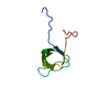 1j3tS S: 精密化の開始モデル |
|---|---|
| 類似構造データ | |
| その他のデータベース |

-リンク
 PDBj
PDBj


- 集合体
集合体
| 登録構造単位 | 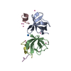
| ||||||||
|---|---|---|---|---|---|---|---|---|---|
| 1 |
| ||||||||
| 2 | 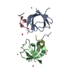
| ||||||||
| 単位格子 |
|
-要素
| #1: タンパク質 | 分子量: 7883.971 Da / 分子数: 2 / 由来タイプ: 組換発現 / 由来: (組換発現) Homo sapiens (ヒト) / 遺伝子: ITSN1, ITSN, SH3D1A / プラスミド: pHH0239 / 発現宿主:  Escherichia coli (大腸菌) / 株 (発現宿主): BL21 / 参照: UniProt: Q15811 Escherichia coli (大腸菌) / 株 (発現宿主): BL21 / 参照: UniProt: Q15811#2: タンパク質・ペプチド | 分子量: 1441.591 Da / 分子数: 3 / 由来タイプ: 合成 / 詳細: synthetic sequence #3: 化合物 | ChemComp-UNX /   分子数: 11 / 由来タイプ: 合成 分子数: 11 / 由来タイプ: 合成#4: 水 | ChemComp-HOH / | 水 分子量: 18.015 Da / 分子数: 99 / 由来タイプ: 天然 / 式: H2O 分子量: 18.015 Da / 分子数: 99 / 由来タイプ: 天然 / 式: H2O |
|---|
-実験情報
-実験
| 実験 | 手法: X線回折 / 使用した結晶の数: 1 |
|---|
- 試料調製
試料調製
| 結晶 | マシュー密度: 1.82 Å3/Da / 溶媒含有率: 32.34 % |
|---|---|
| 結晶化 | 温度: 291 K / 手法: 蒸気拡散法, ハンギングドロップ法 / pH: 8.5 詳細: 1.2 M NaCitrate and 0.1 M Tris, pH 8.5, vapor diffusion hanging drop, temperature 291K |
-データ収集
| 回折 | 平均測定温度: 100 K | |||||||||||||||||||||||||||||||||||||||||||||||||||||||||||||||||||||||||||||||||||||||||||||||||||||||||||||||||||||||||||||||||||||||||||||||||||
|---|---|---|---|---|---|---|---|---|---|---|---|---|---|---|---|---|---|---|---|---|---|---|---|---|---|---|---|---|---|---|---|---|---|---|---|---|---|---|---|---|---|---|---|---|---|---|---|---|---|---|---|---|---|---|---|---|---|---|---|---|---|---|---|---|---|---|---|---|---|---|---|---|---|---|---|---|---|---|---|---|---|---|---|---|---|---|---|---|---|---|---|---|---|---|---|---|---|---|---|---|---|---|---|---|---|---|---|---|---|---|---|---|---|---|---|---|---|---|---|---|---|---|---|---|---|---|---|---|---|---|---|---|---|---|---|---|---|---|---|---|---|---|---|---|---|---|---|---|
| 放射光源 | 由来: 回転陽極 / タイプ: RIGAKU FR-E SUPERBRIGHT / 波長: 1.54178 Å | |||||||||||||||||||||||||||||||||||||||||||||||||||||||||||||||||||||||||||||||||||||||||||||||||||||||||||||||||||||||||||||||||||||||||||||||||||
| 検出器 | タイプ: ADSC QUANTUM 315r / 検出器: CCD / 日付: 2012年7月29日 | |||||||||||||||||||||||||||||||||||||||||||||||||||||||||||||||||||||||||||||||||||||||||||||||||||||||||||||||||||||||||||||||||||||||||||||||||||
| 放射 | プロトコル: SINGLE WAVELENGTH / 単色(M)・ラウエ(L): M / 散乱光タイプ: x-ray | |||||||||||||||||||||||||||||||||||||||||||||||||||||||||||||||||||||||||||||||||||||||||||||||||||||||||||||||||||||||||||||||||||||||||||||||||||
| 放射波長 | 波長: 1.54178 Å / 相対比: 1 | |||||||||||||||||||||||||||||||||||||||||||||||||||||||||||||||||||||||||||||||||||||||||||||||||||||||||||||||||||||||||||||||||||||||||||||||||||
| 反射 | 解像度: 1.8→50 Å / Num. all: 13540 / Num. obs: 13540 / % possible obs: 95.8 % / Observed criterion σ(F): 0 / Observed criterion σ(I): 0 / 冗長度: 7.6 % / Biso Wilson estimate: 23.5 Å2 / Rmerge(I) obs: 0.03 / Χ2: 0.186 / Net I/σ(I): 28.7 | |||||||||||||||||||||||||||||||||||||||||||||||||||||||||||||||||||||||||||||||||||||||||||||||||||||||||||||||||||||||||||||||||||||||||||||||||||
| 反射 シェル |
|
- 解析
解析
| ソフトウェア |
| ||||||||||||||||||||||||||||||||||||||||||||||||||||||||||||
|---|---|---|---|---|---|---|---|---|---|---|---|---|---|---|---|---|---|---|---|---|---|---|---|---|---|---|---|---|---|---|---|---|---|---|---|---|---|---|---|---|---|---|---|---|---|---|---|---|---|---|---|---|---|---|---|---|---|---|---|---|---|
| 精密化 | 構造決定の手法: 分子置換 開始モデル: 1J3T 解像度: 1.8→29.12 Å / Cor.coef. Fo:Fc: 0.963 / Cor.coef. Fo:Fc free: 0.939 / WRfactor Rfree: 0.2231 / WRfactor Rwork: 0.1754 / Occupancy max: 1 / Occupancy min: 0.3 / FOM work R set: 0.8283 / SU B: 3.253 / SU ML: 0.102 / SU R Cruickshank DPI: 0.1465 / SU Rfree: 0.1453 / 交差検証法: THROUGHOUT / σ(F): 0 / ESU R: 0.146 / ESU R Free: 0.145 / 立体化学のターゲット値: MAXIMUM LIKELIHOOD
| ||||||||||||||||||||||||||||||||||||||||||||||||||||||||||||
| 溶媒の処理 | イオンプローブ半径: 0.8 Å / 減衰半径: 0.8 Å / VDWプローブ半径: 1.2 Å / 溶媒モデル: MASK | ||||||||||||||||||||||||||||||||||||||||||||||||||||||||||||
| 原子変位パラメータ | Biso max: 64.26 Å2 / Biso mean: 26.3598 Å2 / Biso min: 12.68 Å2
| ||||||||||||||||||||||||||||||||||||||||||||||||||||||||||||
| 精密化ステップ | サイクル: LAST / 解像度: 1.8→29.12 Å
| ||||||||||||||||||||||||||||||||||||||||||||||||||||||||||||
| 拘束条件 |
| ||||||||||||||||||||||||||||||||||||||||||||||||||||||||||||
| LS精密化 シェル | 解像度: 1.802→1.848 Å / Total num. of bins used: 20
|
