 Movie
Movie Controller


+ Open data

- Basic information
| Entry | Database: PDB / ID: 4coc | ||||||
|---|---|---|---|---|---|---|---|
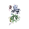
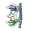
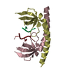
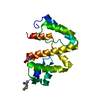
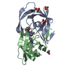
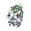
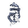
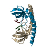
| Title | HIV-1 capsid C-terminal domain mutant (Y169L) | ||||||
 Components Components | CAPSID PROTEIN P24 | ||||||
 Keywords Keywords | VIRAL PROTEIN / VIRUS ASSEMBLY / HELICAL RECONSTRUCTION | ||||||
| Function / homology |  Function and homology information Function and homology informationHIV-1 retropepsin / symbiont-mediated activation of host apoptosis / retroviral ribonuclease H / exoribonuclease H / exoribonuclease H activity / DNA integration / viral genome integration into host DNA / establishment of integrated proviral latency / RNA-directed DNA polymerase / RNA stem-loop binding ...HIV-1 retropepsin / symbiont-mediated activation of host apoptosis / retroviral ribonuclease H / exoribonuclease H / exoribonuclease H activity / DNA integration / viral genome integration into host DNA / establishment of integrated proviral latency / RNA-directed DNA polymerase / RNA stem-loop binding / viral penetration into host nucleus / host multivesicular body / RNA-directed DNA polymerase activity / RNA-DNA hybrid ribonuclease activity / Transferases; Transferring phosphorus-containing groups; Nucleotidyltransferases / host cell / viral nucleocapsid / DNA recombination / DNA-directed DNA polymerase / aspartic-type endopeptidase activity / Hydrolases; Acting on ester bonds / DNA-directed DNA polymerase activity / symbiont-mediated suppression of host gene expression / viral translational frameshifting / symbiont entry into host cell / lipid binding / host cell nucleus / host cell plasma membrane / virion membrane / structural molecule activity / proteolysis / DNA binding / zinc ion binding Similarity search - Function | ||||||
| Biological species |   HUMAN IMMUNODEFICIENCY VIRUS 1 HUMAN IMMUNODEFICIENCY VIRUS 1 | ||||||
| Method |  X-RAY DIFFRACTION / SYNCHROTRON / MOLECULAR REPLACEMENT / Resolution: 1.59 Å X-RAY DIFFRACTION / SYNCHROTRON / MOLECULAR REPLACEMENT / Resolution: 1.59 Å | ||||||
 Authors Authors | Bharat, T.A.M. / Castillo-Menendez, L.R. / Hagen, W.J.H. / Lux, V. / Igonet, S. / Schorb, M. / Schur, F.K.M. / Krausslich, H.-G. / Briggs, J.A.G. | ||||||
 Citation Citation | Journal: Proc Natl Acad Sci U S A / Year: 2014 Title: Cryo-electron microscopy of tubular arrays of HIV-1 Gag resolves structures essential for immature virus assembly. Authors: Tanmay A M Bharat / Luis R Castillo Menendez / Wim J H Hagen / Vanda Lux / Sebastien Igonet / Martin Schorb / Florian K M Schur / Hans-Georg Kräusslich / John A G Briggs /    Abstract: The assembly of HIV-1 is mediated by oligomerization of the major structural polyprotein, Gag, into a hexameric protein lattice at the plasma membrane of the infected cell. This leads to budding and ...The assembly of HIV-1 is mediated by oligomerization of the major structural polyprotein, Gag, into a hexameric protein lattice at the plasma membrane of the infected cell. This leads to budding and release of progeny immature virus particles. Subsequent proteolytic cleavage of Gag triggers rearrangement of the particles to form mature infectious virions. Obtaining a structural model of the assembled lattice of Gag within immature virus particles is necessary to understand the interactions that mediate assembly of HIV-1 particles in the infected cell, and to describe the substrate that is subsequently cleaved by the viral protease. An 8-Å resolution structure of an immature virus-like tubular array assembled from a Gag-derived protein of the related retrovirus Mason-Pfizer monkey virus (M-PMV) has previously been reported, and a model for the arrangement of the HIV-1 capsid (CA) domains has been generated based on homology to this structure. Here we have assembled tubular arrays of a HIV-1 Gag-derived protein with an immature-like arrangement of the C-terminal CA domains and have solved their structure by using hybrid cryo-EM and tomography analysis. The structure reveals the arrangement of the C-terminal domain of CA within an immature-like HIV-1 Gag lattice, and provides, to our knowledge, the first high-resolution view of the region immediately downstream of CA, which is essential for assembly, and is significantly different from the respective region in M-PMV. Our results reveal a hollow column of density for this region in HIV-1 that is compatible with the presence of a six-helix bundle at this position. | ||||||
| History |
|
- Structure visualization
| Structure viewer | Molecule: MolmilJmol/JSmol |
|---|
- Downloads & links
-Download
| PDBx/mmCIF format | 4coc.cif.gz | 61 KB | Display | PDBx/mmCIF format |
|---|---|---|---|---|
| PDB format | pdb4coc.ent.gz | 45.3 KB | Display | PDB format |
| PDBx/mmJSON format | 4coc.json.gz | Tree view | PDBx/mmJSON format | |
| Others |  Other downloads Other downloads |
-Validation report
| Arichive directory | https://data.pdbj.org/pub/pdb/validation_reports/co/4cocftp://data.pdbj.org/pub/pdb/validation_reports/co/4coc | HTTPS FTP |
|---|
-Related structure data
| Related structure data |  2638C 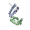 4copC 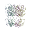 4d1kC  2buoS S: Starting model for refinement C: citing same article ( |
|---|---|
| Similar structure data |


-Links
 PDBj
PDBj
- Assembly
| Deposited unit | 
| ||||||||
|---|---|---|---|---|---|---|---|---|---|
| 1 | 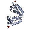
| ||||||||
| 2 | 
| ||||||||
| 3 | 
| ||||||||
| Unit cell |
|
-Components
| #1: Protein | Mass: 9480.904 Da / Num. of mol.: 3 / Fragment: C-TERMINAL DOMAIN, RESIDUES 278-363 / Mutation: YES Source method: isolated from a genetically manipulated source Source: (gene. exp.) HUMAN IMMUNODEFICIENCY VIRUS 1 / Strain: NL4-3 / Production host:  #2: Chemical |   Mass: 96.063 Da / Num. of mol.: 2 / Source method: obtained synthetically / Formula: SO4 Mass: 96.063 Da / Num. of mol.: 2 / Source method: obtained synthetically / Formula: SO4#3: Water | ChemComp-HOH / |  Mass: 18.015 Da / Num. of mol.: 174 / Source method: isolated from a natural source / Formula: H2O Mass: 18.015 Da / Num. of mol.: 174 / Source method: isolated from a natural source / Formula: H2O |
|---|
-Experimental details
-Experiment
| Experiment | Method: X-RAY DIFFRACTION / Number of used crystals: 1 |
|---|
- Sample preparation
| Crystal | Density Matthews: 2.47 Å3/Da / Density % sol: 50.28 % / Description: NONE |
|---|---|
| Crystal grow | pH: 8.5 / Details: 30% PEG 4000, 0.2 M LISO4, 0.1 M TRIS PH 8.5 |
-Data collection
| Diffraction | Mean temperature: 100 K |
|---|---|
| Diffraction source | Source: SYNCHROTRON / Site: ESRF / Beamline: ID23-2 / Wavelength: 0.873 |
| Detector | Type: MARMOSAIC 225 mm CCD / Detector: CCD Details: ONE PAIR OF (300X40X15) MM3 LONG PT COATED SI MIRROR, 260MM USABLE, IN A KIRKPATRICK-BAEZ GEOMETRY |
| Radiation | Monochromator: HORIZONTALLY SIDE DIFFRACTING SILICON 111 CRYSTAL Protocol: SINGLE WAVELENGTH / Monochromatic (M) / Laue (L): M / Scattering type: x-ray |
| Radiation wavelength | Wavelength: 0.873 Å / Relative weight: 1 |
| Reflection | Resolution: 1.59→47.42 Å / Num. obs: 33062 / % possible obs: 97.9 % / Observed criterion σ(I): 2 / Redundancy: 5.6 % / Rmerge(I) obs: 0.09 / Net I/σ(I): 13.1 |
| Reflection shell | Resolution: 1.59→1.68 Å / Redundancy: 2.6 % / Rmerge(I) obs: 0.52 / Mean I/σ(I) obs: 2 / % possible all: 77.8 |
- Processing
| Software |
| ||||||||||||||||||||||||||||||||||||||||||||||||||||||||||||||||||||||||||||||||||||||||||||||||||||||||||||||||||||||||||||||||||||||||||||||||||||||||||||||||||||||||||||||||||||||
|---|---|---|---|---|---|---|---|---|---|---|---|---|---|---|---|---|---|---|---|---|---|---|---|---|---|---|---|---|---|---|---|---|---|---|---|---|---|---|---|---|---|---|---|---|---|---|---|---|---|---|---|---|---|---|---|---|---|---|---|---|---|---|---|---|---|---|---|---|---|---|---|---|---|---|---|---|---|---|---|---|---|---|---|---|---|---|---|---|---|---|---|---|---|---|---|---|---|---|---|---|---|---|---|---|---|---|---|---|---|---|---|---|---|---|---|---|---|---|---|---|---|---|---|---|---|---|---|---|---|---|---|---|---|---|---|---|---|---|---|---|---|---|---|---|---|---|---|---|---|---|---|---|---|---|---|---|---|---|---|---|---|---|---|---|---|---|---|---|---|---|---|---|---|---|---|---|---|---|---|---|---|---|---|
| Refinement | Method to determine structure: MOLECULAR REPLACEMENT Starting model: PDB ENTRY 2BUO Resolution: 1.59→47.42 Å / Cor.coef. Fo:Fc: 0.958 / Cor.coef. Fo:Fc free: 0.948 / SU B: 1.682 / SU ML: 0.059 / Cross valid method: THROUGHOUT / ESU R: 0.088 / ESU R Free: 0.091 / Stereochemistry target values: MAXIMUM LIKELIHOOD Details: HYDROGENS HAVE BEEN ADDED IN THE RIDING POSITIONS. U VALUES HAVE BEEN REFINED INDIVIDUALLY
| ||||||||||||||||||||||||||||||||||||||||||||||||||||||||||||||||||||||||||||||||||||||||||||||||||||||||||||||||||||||||||||||||||||||||||||||||||||||||||||||||||||||||||||||||||||||
| Solvent computation | Ion probe radii: 0.8 Å / Shrinkage radii: 0.8 Å / VDW probe radii: 1.2 Å / Solvent model: MASK | ||||||||||||||||||||||||||||||||||||||||||||||||||||||||||||||||||||||||||||||||||||||||||||||||||||||||||||||||||||||||||||||||||||||||||||||||||||||||||||||||||||||||||||||||||||||
| Displacement parameters | Biso mean: 22.288 Å2
| ||||||||||||||||||||||||||||||||||||||||||||||||||||||||||||||||||||||||||||||||||||||||||||||||||||||||||||||||||||||||||||||||||||||||||||||||||||||||||||||||||||||||||||||||||||||
| Refinement step | Cycle: LAST / Resolution: 1.59→47.42 Å
| ||||||||||||||||||||||||||||||||||||||||||||||||||||||||||||||||||||||||||||||||||||||||||||||||||||||||||||||||||||||||||||||||||||||||||||||||||||||||||||||||||||||||||||||||||||||
| Refine LS restraints |
|