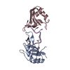
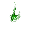
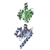
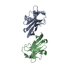
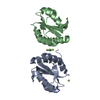
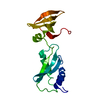
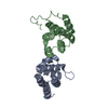
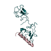
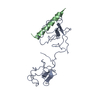
登録情報 データベース : PDB / ID : 2doiタイトル The X-ray crystallographic structure of the angiogenesis inhibitor, angiostatin, bound to a peptide from the group A streptococcus protein PAM Angiostatin Plasminogen-binding group A streptococcal M-like protein PAM キーワード / / / / 機能・相同性 分子機能 ドメイン・相同性 構成要素
/ / / / / / / / / / / / / / / / / / / / / / / / / / / / / / / / / / / / / / / / / / / / / / / / / / / / / / / / / / / / / / / / / / / / / / / / / / / / / / / / / / / / / / / / / / / / / / / / / / / 生物種 Homo sapiens (ヒト)手法 / / / 解像度 : 3.1 Å データ登録者 Cnudde, S.E. / Prorok, M. / Castellino, F.J. / Geiger, J.H. ジャーナル : Biochemistry / 年 : 2006タイトル : X-ray crystallographic structure of the angiogenesis inhibitor, angiostatin, bound to a peptide from the group A streptococcal surface protein PAM著者 : Cnudde, S.E. / Prorok, M. / Castellino, F.J. / Geiger, J.H. 履歴 登録 2006年4月29日 登録サイト / 処理サイト 改定 1.0 2006年12月5日 Provider / タイプ 改定 1.1 2008年4月30日 Group 改定 1.2 2011年7月13日 Group / Version format compliance改定 1.3 2017年10月11日 Group / カテゴリ Item _software.classification / _software.contact_author ... _software.classification / _software.contact_author / _software.contact_author_email / _software.date / _software.language / _software.location / _software.name / _software.type / _software.version 改定 1.4 2021年11月10日 Group / カテゴリ / struct_ref_seq_difItem / _database_2.pdbx_database_accession / _struct_ref_seq_dif.details改定 1.5 2023年10月25日 Group / Refinement descriptionカテゴリ / chem_comp_bond / pdbx_initial_refinement_model
すべて表示 表示を減らす
 ムービー
ムービー コントローラー


 データを開く
 基本情報
基本情報 要素
要素 キーワード
キーワード HYDROLASE (加水分解酵素) /
  機能・相同性情報
 データ登録者
 引用
引用 構造の表示
構造の表示 ダウンロードとリンク
 その他のダウンロード
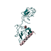


 PDBj
PDBj


 集合体
集合体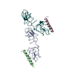

 試料調製
試料調製 / ビームライン: 5ID-B / 波長: 1.0719 Å
 解析
解析