ムービー
ムービー コントローラー


+ データを開く

- 基本情報
基本情報
| 登録情報 | データベース: PDB / ID: 1t1o | ||||||
|---|---|---|---|---|---|---|---|
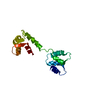
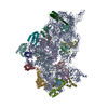
| タイトル | Components of the control 70S ribosome to provide reference for the RRF binding site | ||||||
 要素 要素 |
| ||||||
 キーワード キーワード |  RIBOSOME (リボソーム) / RRF binding position on the ribosome RIBOSOME (リボソーム) / RRF binding position on the ribosome | ||||||
| 機能・相同性 | リボ核酸 / RNA (> 10) 機能・相同性情報 機能・相同性情報 | ||||||
| 生物種 |  Escherichia coli (大腸菌) Escherichia coli (大腸菌) | ||||||
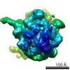
| 手法 | 電子顕微鏡法 / 単粒子再構成法 / クライオ電子顕微鏡法 / 解像度: 12 Å | ||||||
 データ登録者 データ登録者 | Agrawal, R.K. / Sharma, M.R. / Kiel, M.C. / Hirokawa, G. / Booth, T.M. / Spahn, C.M. / Grassucci, R.A. / Kaji, A. / Frank, J. | ||||||
 引用 引用 | ジャーナル: Proc Natl Acad Sci U S A / 年: 2004 タイトル: Visualization of ribosome-recycling factor on the Escherichia coli 70S ribosome: functional implications. 著者: Rajendra K Agrawal / Manjuli R Sharma / Michael C Kiel / Go Hirokawa / Timothy M Booth / Christian M T Spahn / Robert A Grassucci / Akira Kaji / Joachim Frank /  要旨: After the termination step of protein synthesis, a deacylated tRNA and mRNA remain associated with the ribosome. The ribosome-recycling factor (RRF), together with elongation factor G (EF-G), ...After the termination step of protein synthesis, a deacylated tRNA and mRNA remain associated with the ribosome. The ribosome-recycling factor (RRF), together with elongation factor G (EF-G), disassembles this posttermination complex into mRNA, tRNA, and the ribosome. We have obtained a three-dimensional cryo-electron microscopic map of a complex of the Escherichia coli 70S ribosome and RRF. We find that RRF interacts mainly with the segments of the large ribosomal subunit's (50S) rRNA helices that are involved in the formation of two central intersubunit bridges, B2a and B3. The binding of RRF induces considerable conformational changes in some of the functional domains of the ribosome. As compared to its binding position derived previously by hydroxyl radical probing study, we find that RRF binds further inside the intersubunit space of the ribosome such that the tip of its domain I is shifted (by approximately 13 A) toward protein L5 within the central protuberance of the 50S subunit, and domain II is oriented more toward the small ribosomal subunit (30S). Overlapping binding sites of RRF, EF-G, and the P-site tRNA suggest that the binding of EF-G would trigger the removal of deacylated tRNA from the P site by moving RRF toward the ribosomal E site, and subsequent removal of mRNA may be induced by a shift in the position of 16S rRNA helix 44, which harbors part of the mRNA. #1: ジャーナル: Cell / 年: 2001タイトル: High resolution structure of the large ribosomal subunit from a mesophilic eubacterium. 著者: J Harms / F Schluenzen / R Zarivach / A Bashan / S Gat / I Agmon / H Bartels / F Franceschi / A Yonath /  要旨: We describe the high resolution structure of the large ribosomal subunit from Deinococcus radiodurans (D50S), a gram-positive mesophile suitable for binding of antibiotics and functionally relevant ...We describe the high resolution structure of the large ribosomal subunit from Deinococcus radiodurans (D50S), a gram-positive mesophile suitable for binding of antibiotics and functionally relevant ligands. The over-all structure of D50S is similar to that from the archae bacterium Haloarcula marismortui (H50S); however, a detailed comparison revealed significant differences, for example, in the orientation of nucleotides in peptidyl transferase center and in the structures of many ribosomal proteins. Analysis of ribosomal features involved in dynamic aspects of protein biosynthesis that are partially or fully disordered in H50S revealed the conformations of intersubunit bridges in unbound subunits, suggesting how they may change upon subunit association and how movements of the L1-stalk may facilitate the exit of tRNA. #2: ジャーナル: Nature / 年: 2000タイトル: Structure of the 30S ribosomal subunit. 著者: B T Wimberly / D E Brodersen / W M Clemons / R J Morgan-Warren / A P Carter / C Vonrhein / T Hartsch / V Ramakrishnan /  要旨: Genetic information encoded in messenger RNA is translated into protein by the ribosome, which is a large nucleoprotein complex comprising two subunits, denoted 30S and 50S in bacteria. Here we ...Genetic information encoded in messenger RNA is translated into protein by the ribosome, which is a large nucleoprotein complex comprising two subunits, denoted 30S and 50S in bacteria. Here we report the crystal structure of the 30S subunit from Thermus thermophilus, refined to 3 A resolution. The final atomic model rationalizes over four decades of biochemical data on the ribosome, and provides a wealth of information about RNA and protein structure, protein-RNA interactions and ribosome assembly. It is also a structural basis for analysis of the functions of the 30S subunit, such as decoding, and for understanding the action of antibiotics. The structure will facilitate the interpretation in molecular terms of lower resolution structural data on several functional states of the ribosome from electron microscopy and crystallography. | ||||||
| 履歴 |
|
- 構造の表示
構造の表示
| ムービー |
ムービービューア |
|---|---|
| 構造ビューア | 分子: MolmilJmol/JSmol |
- ダウンロードとリンク
-ダウンロード
| PDBx/mmCIF形式 | 1t1o.cif.gz | 9.9 KB | 表示 | PDBx/mmCIF形式 |
|---|---|---|---|---|
| PDB形式 | pdb1t1o.ent.gz | 4.7 KB | 表示 | PDB形式 |
| PDBx/mmJSON形式 | 1t1o.json.gz | ツリー表示 | PDBx/mmJSON形式 | |
| その他 |  その他のダウンロード その他のダウンロード |
-検証レポート
| アーカイブディレクトリ | https://data.pdbj.org/pub/pdb/validation_reports/t1/1t1oftp://data.pdbj.org/pub/pdb/validation_reports/t1/1t1o | HTTPS FTP |
|---|
-関連構造データ

-リンク
 PDBj
PDBj


- 集合体
集合体
| 登録構造単位 | 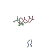
|
|---|---|
| 1 |
|
-要素
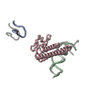
| #1: RNA鎖 | 分子量: 3852.344 Da / 分子数: 1 / 断片: Apical loop of Helix 43 / 由来タイプ: 天然 / 詳細: fitted into the cryo-EM map of the 70S ribosome / 由来: (天然) Escherichia coli (大腸菌) / 株: MRE600 |
|---|---|
| #2: RNA鎖 | 分子量: 6077.673 Da / 分子数: 1 / 断片: Helix 69 / 由来タイプ: 天然 / 詳細: fitted into the cryo-EM map of the 70S ribosome / 由来: (天然) Escherichia coli (大腸菌) / 株: MRE600 |
| #3: RNA鎖 | 分子量: 13581.124 Da / 分子数: 1 / 断片: Top portion of helix 44 / 由来タイプ: 天然 / 詳細: fitted into the cryo-EM map of the 70S ribosome / 由来: (天然) Escherichia coli (大腸菌) / 株: MRE600 |
-実験情報
-実験
| 実験 | 手法: 電子顕微鏡法 |
|---|---|
| EM実験 | 試料の集合状態: PARTICLE / 3次元再構成法: 単粒子再構成法 |
- 試料調製
試料調製
| 構成要素 | 名称: 70S-RRF complex / タイプ: RIBOSOME |
|---|---|
| 緩衝液 | pH: 7.5 |
| 試料 | 濃度: 32 mg/ml / 包埋: NO / シャドウイング: NO / 染色: NO / 凍結: YES |
| 試料支持 | 詳細: Quantifoil holley-carbon film grids |
| 急速凍結 | 装置: HOMEMADE PLUNGER / 凍結剤: ETHANE / 詳細: Rapid-freezing in liquid ethane |
- 電子顕微鏡撮影
| 実験機器 |  モデル: Tecnai F20 / 画像提供: FEI Company |
|---|---|
| 顕微鏡 | モデル: FEI TECNAI F20 / 日付: 2002年6月1日 |
| 電子銃 | 電子線源: FIELD EMISSION GUN / 加速電圧: 200 kV / 照射モード: FLOOD BEAM |
| 電子レンズ | モード: BRIGHT FIELDBright-field microscopy / 倍率(公称値): 50000 X / 倍率(補正後): 49696 X / 最大 デフォーカス(公称値): 4400 nm / 最小 デフォーカス(公称値): 1400 nm / Cs: 2 mm |
| 試料ホルダ | 温度: 93 K / 傾斜角・最大: 0 ° / 傾斜角・最小: 0 ° |
| 撮影 | 電子線照射量: 20 e/Å2 / フィルム・検出器のモデル: KODAK SO-163 FILM |
- 解析
解析
| CTF補正 | 詳細: CTF correction of 3D-maps by Wiener filtration | |||||||||||||||||||||
|---|---|---|---|---|---|---|---|---|---|---|---|---|---|---|---|---|---|---|---|---|---|---|
| 対称性 | 点対称性: C1 (非対称) | |||||||||||||||||||||
| 3次元再構成 | 手法: 3D projection matching; conjugate gradients with regularization 解像度: 12 Å / 粒子像の数: 51217 / ピクセルサイズ(実測値): 2.82 Å / 倍率補正: TMV / 詳細: SPIDER package / 対称性のタイプ: POINT | |||||||||||||||||||||
| 原子モデル構築 | プロトコル: OTHER 詳細: METHOD--Cross corelation coefficient based manual fitting in O | |||||||||||||||||||||
| 原子モデル構築 |
| |||||||||||||||||||||
| 精密化ステップ | サイクル: LAST
|