 ムービー
ムービー コントローラー


+ データを開く

- 基本情報
基本情報
| 登録情報 | データベース: EMDB / ID: EMD-4486 | |||||||||
|---|---|---|---|---|---|---|---|---|---|---|
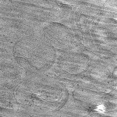
| タイトル | Correlative FM and cryo-ET of GFP-Bax in HeLa cells | |||||||||
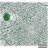
 マップデータ マップデータ | Reconstructed electron cryo-tomogram of HeLa cell overexpressing GFP-Bax | |||||||||
 試料 試料 |
| |||||||||
| 生物種 |  Homo sapiens (ヒト) Homo sapiens (ヒト) | |||||||||
| 手法 | 電子線トモグラフィー法 / クライオ電子顕微鏡法 | |||||||||
 データ登録者 データ登録者 | Ader NR / Hoffmann PC / Ganeva I / Borgeaud AC / Wang C / Youle RJ / Kukulski W | |||||||||
 引用 引用 | ジャーナル: Elife / 年: 2019 タイトル: Molecular and topological reorganizations in mitochondrial architecture interplay during Bax-mediated steps of apoptosis. 著者: Nicholas R Ader / Patrick C Hoffmann / Iva Ganeva / Alicia C Borgeaud / Chunxin Wang / Richard J Youle / Wanda Kukulski /   要旨: During apoptosis, Bcl-2 proteins such as Bax and Bak mediate the release of pro-apoptotic proteins from the mitochondria by clustering on the outer mitochondrial membrane and thereby permeabilizing ...During apoptosis, Bcl-2 proteins such as Bax and Bak mediate the release of pro-apoptotic proteins from the mitochondria by clustering on the outer mitochondrial membrane and thereby permeabilizing it. However, it remains unclear how outer membrane openings form. Here, we combined different correlative microscopy and electron cryo-tomography approaches to visualize the effects of Bax activity on mitochondria in human cells. Our data show that Bax clusters localize near outer membrane ruptures of highly variable size. Bax clusters contain structural elements suggesting a higher order organization of their components. Furthermore, unfolding of inner membrane cristae is coupled to changes in the supramolecular assembly of ATP synthases, particularly pronounced at membrane segments exposed to the cytosol by ruptures. Based on our results, we propose a comprehensive model in which molecular reorganizations of the inner membrane and sequestration of outer membrane components into Bax clusters interplay in the formation of outer membrane ruptures. EDITORIAL NOTE: This article has been through an editorial process in which the authors decide how to respond to the issues raised during peer review. The Reviewing Editor's assessment is that all ...EDITORIAL NOTE: This article has been through an editorial process in which the authors decide how to respond to the issues raised during peer review. The Reviewing Editor's assessment is that all the issues have been addressed (see decision letter). | |||||||||
| 履歴 |
|
- 構造の表示
構造の表示
| ムービー |
 ムービービューア ムービービューア |
|---|---|
| 添付画像 |
- ダウンロードとリンク
-EMDBアーカイブ
| マップデータ | emd_4486.map.gz | 894 MB | EMDBマップデータ形式 | |
|---|---|---|---|---|
| ヘッダ (付随情報) | emd-4486-v30.xmlemd-4486.xml | 12.5 KB 12.5 KB | 表示 表示 | EMDBヘッダ |
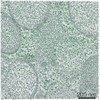
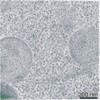
| 画像 |  emd_4486.png emd_4486.png | 139.7 KB | ||
| アーカイブディレクトリ |  http://ftp.pdbj.org/pub/emdb/structures/EMD-4486ftp://ftp.pdbj.org/pub/emdb/structures/EMD-4486 http://ftp.pdbj.org/pub/emdb/structures/EMD-4486ftp://ftp.pdbj.org/pub/emdb/structures/EMD-4486 | HTTPS FTP |
-検証レポート
| 文書・要旨 | emd_4486_validation.pdf.gz | 163.2 KB | 表示 | EMDB検証レポート |
|---|---|---|---|---|
| 文書・詳細版 | emd_4486_full_validation.pdf.gz | 162.3 KB | 表示 | |
| XML形式データ | emd_4486_validation.xml.gz | 5 KB | 表示 | |
| アーカイブディレクトリ | https://ftp.pdbj.org/pub/emdb/validation_reports/EMD-4486ftp://ftp.pdbj.org/pub/emdb/validation_reports/EMD-4486 | HTTPS FTP |
-関連構造データ
-リンク
| EMDBのページ | EMDB (EBI/PDBe) / EMDataResource |
|---|
-マップ
| ファイル | ダウンロード / ファイル: emd_4486.map.gz / 形式: CCP4 / 大きさ: 1.3 GB / タイプ: IMAGE STORED AS SIGNED BYTE | ||||||||||||||||||||||||||||||||||||||||||||||||||||||||||||
|---|---|---|---|---|---|---|---|---|---|---|---|---|---|---|---|---|---|---|---|---|---|---|---|---|---|---|---|---|---|---|---|---|---|---|---|---|---|---|---|---|---|---|---|---|---|---|---|---|---|---|---|---|---|---|---|---|---|---|---|---|---|
| 注釈 | Reconstructed electron cryo-tomogram of HeLa cell overexpressing GFP-Bax | ||||||||||||||||||||||||||||||||||||||||||||||||||||||||||||
| ボクセルのサイズ | X=Y=Z: 7.534 Å | ||||||||||||||||||||||||||||||||||||||||||||||||||||||||||||
| 密度 |
| ||||||||||||||||||||||||||||||||||||||||||||||||||||||||||||
| 対称性 | 空間群: 1 | ||||||||||||||||||||||||||||||||||||||||||||||||||||||||||||
| 詳細 | EMDB XML:
CCP4マップ ヘッダ情報:
| ||||||||||||||||||||||||||||||||||||||||||||||||||||||||||||
-添付データ
- 試料の構成要素
-全体 : HeLa (homo sapiens)
| 全体 | 名称: HeLa (homo sapiens) |
|---|---|
| 要素 |
|
-超分子 #1: HeLa (homo sapiens)
| 超分子 | 名称: HeLa (homo sapiens) / タイプ: cell / ID: 1 / 親要素: 0 詳細: Cryofixation of cell was performed 16 h after transfection of GFP-Bax plasmid. |
|---|---|
| 由来(天然) | 生物種: Homo sapiens (ヒト) |
-実験情報
-構造解析
| 手法 | クライオ電子顕微鏡法 |
|---|---|
 解析 解析 | 電子線トモグラフィー法 |
| 試料の集合状態 | cell |
-試料調製
| 緩衝液 | pH: 7.4 / 構成要素 - 名称: PBS 詳細: DMEM, high glucose, GlutaMAX, pyruvate (Thermo 31996) medium supplemented with 10% heat-inactivated FBS (Gibco 10270), 10 mM HEPES, and 1x NEAA (Thermo 11140) |
|---|---|
| グリッド | モデル: Quantifoil R3.5/1 / 材質: COPPER / 支持フィルム - 材質: CARBON / 支持フィルム - トポロジー: HOLEY / 前処理 - タイプ: PLASMA CLEANING |
| 凍結 | 凍結剤: NITROGEN / 詳細: high pressure frozen. |
| 加圧凍結法 | 装置: OTHER 詳細: HeLa cells were grown for 24 h in 6-well plates, transfected with 2000 ng hBax-C3-EGFP plasmid and incubated with Q-VD-OPh for 16 h, then trypsinized and pelleted. Immediately before ...詳細: HeLa cells were grown for 24 h in 6-well plates, transfected with 2000 ng hBax-C3-EGFP plasmid and incubated with Q-VD-OPh for 16 h, then trypsinized and pelleted. Immediately before trypsinizing, cells were stained with MitoTracker Deep Red. Pellets were maintained at 37 degrees C while they were mixed 1:1 with 40% Dextran (Sigma) in PBS, pipetted into the 0.2 mm recess of gold-coated copper carriers, covered with the flat side of Aluminum carriers B and high-pressure frozen with a Leica HPM100 (Leica Microsystems).. The value given for _emd_high_pressure_freezing.instrument is Leica EM HP100. This is not in a list of allowed values set(['LEICA EM PACT2', 'LEICA EM PACT', 'EMS-002 RAPID IMMERSION FREEZER', 'OTHER', 'LEICA EM HPM100', 'BAL-TEC HPM 010']) so OTHER is written into the XML file. |
| Cryo protectant | 40% Dextran |
| 切片作成 | ウルトラミクロトーム - 装置: Leica UC6/FC6 / ウルトラミクロトーム - 温度: 123 K / ウルトラミクロトーム - 最終 厚さ: 100 nm ウルトラミクロトーム - 詳細: 100 nm thick vitreous sections were produced at -150 degrees C in a UC6/FC6 cryo-ultramicrotome (Leica Microsystems) using cryotrim 25 and a 35 degree cryo ...ウルトラミクロトーム - 詳細: 100 nm thick vitreous sections were produced at -150 degrees C in a UC6/FC6 cryo-ultramicrotome (Leica Microsystems) using cryotrim 25 and a 35 degree cryo immuno knives (Diatome). The sections were attached using a Crion antistatic device (Leica Microsystems) to EM grids (R3.5/1, copper, Quantifoil) that were plasma cleaned and had 100 nm TetraSpeck beads (Invitrogen) diluted 1:50 in PBS adhered to them. |
- 電子顕微鏡法
| 顕微鏡 | FEI TITAN KRIOS |
|---|---|
| 特殊光学系 | エネルギーフィルター - 名称: GIF Quantum LS / エネルギーフィルター - スリット幅: 20 eV |
| 詳細 | Montaged images of the entire grid were acquired at low magnification at pixel size of 182.3 nm. Intermediate magnification maps of grid squares with vitreous sections were acquired at pixel size 5.5 nm. |
| 撮影 | フィルム・検出器のモデル: GATAN K2 SUMMIT (4k x 4k) 検出モード: COUNTING / デジタル化 - サイズ - 横: 3710 pixel / デジタル化 - サイズ - 縦: 3838 pixel / 平均電子線量: 1.1 e/Å2 詳細: Electron cryo-tomographic tilt-series were collected on a Titan Krios (FEI) operated at 300 kV using a Quantum energy filter (slit width 20 eV) and a K2 direct electron detector (Gatan) in ...詳細: Electron cryo-tomographic tilt-series were collected on a Titan Krios (FEI) operated at 300 kV using a Quantum energy filter (slit width 20 eV) and a K2 direct electron detector (Gatan) in counting mode at a pixel size of 3.7 angstroms and at a dose rate of ~ 2-4 e-/pixel/second on the detector, dependent on sample thickness. Tilt-series were acquired between +/- 60 degrees starting from 0 degrees with 1 degrees increment using SerialEM (Mastronarde, 2005) following a grouped dose-symmetric acquisition with a group size of 4 (Bharat et al., 2018; Hagen et al., 2017), and at -5 um defocus. A dose of approximately 1.0 to 1.2 e-/square angstroms was applied per image of the tilt-series. |
| 電子線 | 加速電圧: 300 kV / 電子線源:  FIELD EMISSION GUN FIELD EMISSION GUN |
| 電子光学系 | 照射モード: FLOOD BEAM / 撮影モード: BRIGHT FIELD / 最大 デフォーカス(公称値): 5.0 µm / 最小 デフォーカス(公称値): 5.0 µm |
| 試料ステージ | 試料ホルダーモデル: FEI TITAN KRIOS AUTOGRID HOLDER ホルダー冷却材: NITROGEN |
| 実験機器 |  モデル: Titan Krios / 画像提供: FEI Company |
-画像解析
| 最終 再構成 | アルゴリズム: SIMULTANEOUS ITERATIVE (SIRT) / ソフトウェア - 名称: eTomo (ver. 4.10.20) 詳細: NUmber of tilted images used in for this volume is approximate. 使用した粒子像数: 70 |
|---|
