 Movie
Movie Controller


+ Open data

- Basic information
| Entry | Database: EMDB / ID: EMD-4483 | |||||||||
|---|---|---|---|---|---|---|---|---|---|---|
| Title | Correlative FM and ET of GFP-Bax in HeLa cells | |||||||||
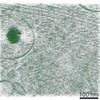
 Map data Map data | Reconstructed tomogram of mitochondria in HeLa cells overexpressing GFP-Bax. | |||||||||
 Sample Sample |
| |||||||||
| Biological species |  Homo sapiens (human) Homo sapiens (human) | |||||||||
| Method | electron tomography / negative staining | |||||||||
 Authors Authors | Ader NR / Hoffmann PC / Ganeva I / Borgeaud AC / Wang C / Youle RJ / Kukulski W | |||||||||
 Citation Citation | Journal: Elife / Year: 2019 Title: Molecular and topological reorganizations in mitochondrial architecture interplay during Bax-mediated steps of apoptosis. Authors: Nicholas R Ader / Patrick C Hoffmann / Iva Ganeva / Alicia C Borgeaud / Chunxin Wang / Richard J Youle / Wanda Kukulski /   Abstract: During apoptosis, Bcl-2 proteins such as Bax and Bak mediate the release of pro-apoptotic proteins from the mitochondria by clustering on the outer mitochondrial membrane and thereby permeabilizing ...During apoptosis, Bcl-2 proteins such as Bax and Bak mediate the release of pro-apoptotic proteins from the mitochondria by clustering on the outer mitochondrial membrane and thereby permeabilizing it. However, it remains unclear how outer membrane openings form. Here, we combined different correlative microscopy and electron cryo-tomography approaches to visualize the effects of Bax activity on mitochondria in human cells. Our data show that Bax clusters localize near outer membrane ruptures of highly variable size. Bax clusters contain structural elements suggesting a higher order organization of their components. Furthermore, unfolding of inner membrane cristae is coupled to changes in the supramolecular assembly of ATP synthases, particularly pronounced at membrane segments exposed to the cytosol by ruptures. Based on our results, we propose a comprehensive model in which molecular reorganizations of the inner membrane and sequestration of outer membrane components into Bax clusters interplay in the formation of outer membrane ruptures. EDITORIAL NOTE: This article has been through an editorial process in which the authors decide how to respond to the issues raised during peer review. The Reviewing Editor's assessment is that all ...EDITORIAL NOTE: This article has been through an editorial process in which the authors decide how to respond to the issues raised during peer review. The Reviewing Editor's assessment is that all the issues have been addressed (see decision letter). | |||||||||
| History |
|
- Structure visualization
| Movie |
 Movie viewer Movie viewer |
|---|---|
| Supplemental images |

- Downloads & links
-EMDB archive
| Map data | emd_4483.map.gz | 646.2 MB | EMDB map data format | |
|---|---|---|---|---|
| Header (meta data) | emd-4483-v30.xmlemd-4483.xml | 11.5 KB 11.5 KB | Display Display | EMDB header |
| Images | 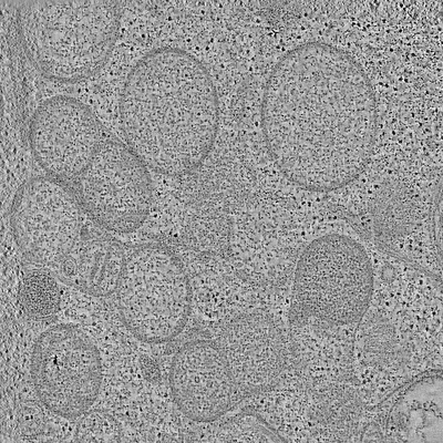 emd_4483.png emd_4483.png | 139.9 KB | ||
| Archive directory |  http://ftp.pdbj.org/pub/emdb/structures/EMD-4483ftp://ftp.pdbj.org/pub/emdb/structures/EMD-4483 http://ftp.pdbj.org/pub/emdb/structures/EMD-4483ftp://ftp.pdbj.org/pub/emdb/structures/EMD-4483 | HTTPS FTP |
-Related structure data

-Links
| EMDB pages | EMDB (EBI/PDBe) / EMDataResource |
|---|
-Map
| File | Download / File: emd_4483.map.gz / Format: CCP4 / Size: 1.3 GB / Type: IMAGE STORED AS SIGNED INTEGER (2 BYTES) | ||||||||||||||||||||||||||||||||||||||||||||||||||||||||||||
|---|---|---|---|---|---|---|---|---|---|---|---|---|---|---|---|---|---|---|---|---|---|---|---|---|---|---|---|---|---|---|---|---|---|---|---|---|---|---|---|---|---|---|---|---|---|---|---|---|---|---|---|---|---|---|---|---|---|---|---|---|---|
| Annotation | Reconstructed tomogram of mitochondria in HeLa cells overexpressing GFP-Bax. | ||||||||||||||||||||||||||||||||||||||||||||||||||||||||||||
| Projections & slices | Image control
Images are generated by Spider. generated in cubic-lattice coordinate | ||||||||||||||||||||||||||||||||||||||||||||||||||||||||||||
| Voxel size | X=Y=Z: 11.02 Å | ||||||||||||||||||||||||||||||||||||||||||||||||||||||||||||
| Density |
| ||||||||||||||||||||||||||||||||||||||||||||||||||||||||||||
| Symmetry | Space group: 1 | ||||||||||||||||||||||||||||||||||||||||||||||||||||||||||||
| Details | EMDB XML:
CCP4 map header:
| ||||||||||||||||||||||||||||||||||||||||||||||||||||||||||||
 Z (Sec.)
 Y (Row.)
 X (Col.)


-Supplemental data
- Sample components
-Entire : HeLa (homo sapiens)
| Entire | Name: HeLa (homo sapiens) |
|---|---|
| Components |
|
-Supramolecule #1: HeLa (homo sapiens)
| Supramolecule | Name: HeLa (homo sapiens) / type: cell / ID: 1 / Parent: 0 Details: Cryofixation of cell was performed 16 h after transfection of GFP-Bax plasmid. |
|---|---|
| Source (natural) | Organism: Homo sapiens (human) |
-Experimental details
-Structure determination
| Method | negative staining |
|---|---|
 Processing Processing | electron tomography |
| Aggregation state | cell |
-Sample preparation
| Buffer | pH: 8.4 / Component - Name: PBS |
|---|---|
| Staining | Type: POSITIVE / Material: Uranyl Acetate / Details: 0.008% uranyl acetate in acetone |
| Sugar embedding | Material: Lowicryl HM20 Details: We use a temperature-controlling Leica AFS2 with FSP. FS is performed at -90 degrees C for 24-36 h in 0.008 percent (w/v) uranyl acetate in glass-distilled acetone. The temperature is then ...Details: We use a temperature-controlling Leica AFS2 with FSP. FS is performed at -90 degrees C for 24-36 h in 0.008 percent (w/v) uranyl acetate in glass-distilled acetone. The temperature is then increased to -45 C (5 degrees C/h). Next, the samples are washed three times with acetone and infiltrated with increasing concentrations (10 percent , 25 percent, 50 percent, 75 percent, 2 h each) of Lowicryl HM20 in acetone. During the final mix, the temperature is raised to -35 degrees C (2.5 degree C/h). The temperature is then raised further to -25 degrees C (2.5 degrees C/h), while 100 percent Lowicryl is exchanged three times in 4 h steps with agitation. Then, UV light is applied for 24 h to initialize Lowicryl polymerization. The temperature is then raised to 20 degrees C (5 degrees C/h). At this point, samples can be taken out of the AFS2, but we wait at least 2 days before removing blocks from the plastic wheel to ensure complete polymerization. |
| Grid | Material: COPPER / Mesh: 200 / Support film - Material: CARBON / Support film - topology: CONTINUOUS |
| High pressure freezing | Instrument: OTHER Details: HPF is accomplished using a Leica HPM100 in the provided temperature- and humidity-controlled chamber. To high-pressure freeze cells, we use a carrier method described in the manual for the ...Details: HPF is accomplished using a Leica HPM100 in the provided temperature- and humidity-controlled chamber. To high-pressure freeze cells, we use a carrier method described in the manual for the Leica EM HPM100 CLEM 3 mm system for HPF of sapphire disks. In brief, carriers are assembled as follows between two plastic half cylinders: (1) 6 mm copper gold-plated support ring in a 6 mm CLEM middle plate, (2) a 3 mm sapphire (cells up), (3) a 3 mm spacer ring, (4) a clean sapphire, and (5) a 6 mm cover ring.. The value given for _emd_high_pressure_freezing.instrument is Leica EM HP100. This is not in a list of allowed values set(['LEICA EM PACT2', 'LEICA EM PACT', 'EMS-002 RAPID IMMERSION FREEZER', 'OTHER', 'LEICA EM HPM100', 'BAL-TEC HPM 010']) so OTHER is written into the XML file. |
| Sectioning | Ultramicrotomy - Instrument: Ultracut E Microtome (Reichert) Ultramicrotomy - Temperature: 296 K / Ultramicrotomy - Final thickness: 300 nm |
| Fiducial marker | Manufacturer: Agar Scientific Ltd. / Diameter: 15 nm |
- Electron microscopy
| Microscope | FEI TECNAI F20 |
|---|---|
| Details | ET was done in STEM mode on an axial brightfield detector with a high-tilt tomography holder (Model 2020; Fischione Instruments) at a 1.1nm pixel size with a camera length of 200 mm. |
| Image recording | Film or detector model: OTHER / Digitization - Dimensions - Width: 2048 pixel / Digitization - Dimensions - Height: 2048 pixel / Average electron dose: 1000.0 e/Å2 Details: ET was done in STEM mode on an axial brightfield detector with a high-tilt tomography holder (Model 2020; Fischione Instruments) at a 1.1nm pixel size with a camera length of 200 mm. [NOTE: ...Details: ET was done in STEM mode on an axial brightfield detector with a high-tilt tomography holder (Model 2020; Fischione Instruments) at a 1.1nm pixel size with a camera length of 200 mm. [NOTE: Electron dose unknown. Value specified here is a dummy] |
| Electron beam | Acceleration voltage: 200 kV / Electron source:  FIELD EMISSION GUN FIELD EMISSION GUN |
| Electron optics | C2 aperture diameter: 50.0 µm / Illumination mode: SPOT SCAN / Imaging mode: BRIGHT FIELD |
| Sample stage | Specimen holder model: OTHER |
| Experimental equipment |  Model: Tecnai F20 / Image courtesy: FEI Company |
-Image processing
| Final reconstruction | Algorithm: BACK PROJECTION / Software - Name: eTomo (ver. 4.10.20) / Details: 237 tilted images used from two axies / Number images used: 237 |
|---|
