 Movie
Movie Controller

[English] 日本語
 Yorodumi
- EMDB-4492: cryo-ET of cryo-FIB milled Bax/Bak double knockout HCT116 cells s... -
+ Open data

- Basic information
| Entry | Database: EMDB / ID: EMD-4492 | |||||||||
|---|---|---|---|---|---|---|---|---|---|---|
| Title | cryo-ET of cryo-FIB milled Bax/Bak double knockout HCT116 cells stably expressing GFP-Bax | |||||||||
 Map data Map data | Reconstructed electron cryo-tomogram of mitochondria in HeLa cells overexpression GFP-Bax | |||||||||
 Sample Sample |
| |||||||||
| Biological species |  Homo sapiens (human) Homo sapiens (human) | |||||||||
| Method | electron tomography / cryo EM | |||||||||
 Authors Authors | Ader NR / Hoffmann PC / Ganeva I / Borgeaud AC / Wang C / Youle RJ / Kukulski W | |||||||||
 Citation Citation | Journal: Elife / Year: 2019 Title: Molecular and topological reorganizations in mitochondrial architecture interplay during Bax-mediated steps of apoptosis. Authors: Nicholas R Ader / Patrick C Hoffmann / Iva Ganeva / Alicia C Borgeaud / Chunxin Wang / Richard J Youle / Wanda Kukulski /   Abstract: During apoptosis, Bcl-2 proteins such as Bax and Bak mediate the release of pro-apoptotic proteins from the mitochondria by clustering on the outer mitochondrial membrane and thereby permeabilizing ...During apoptosis, Bcl-2 proteins such as Bax and Bak mediate the release of pro-apoptotic proteins from the mitochondria by clustering on the outer mitochondrial membrane and thereby permeabilizing it. However, it remains unclear how outer membrane openings form. Here, we combined different correlative microscopy and electron cryo-tomography approaches to visualize the effects of Bax activity on mitochondria in human cells. Our data show that Bax clusters localize near outer membrane ruptures of highly variable size. Bax clusters contain structural elements suggesting a higher order organization of their components. Furthermore, unfolding of inner membrane cristae is coupled to changes in the supramolecular assembly of ATP synthases, particularly pronounced at membrane segments exposed to the cytosol by ruptures. Based on our results, we propose a comprehensive model in which molecular reorganizations of the inner membrane and sequestration of outer membrane components into Bax clusters interplay in the formation of outer membrane ruptures. EDITORIAL NOTE: This article has been through an editorial process in which the authors decide how to respond to the issues raised during peer review. The Reviewing Editor's assessment is that all ...EDITORIAL NOTE: This article has been through an editorial process in which the authors decide how to respond to the issues raised during peer review. The Reviewing Editor's assessment is that all the issues have been addressed (see decision letter). | |||||||||
| History |
|
- Structure visualization
| Movie |
 Movie viewer Movie viewer |
|---|---|
| Supplemental images |

- Downloads & links
-EMDB archive
| Map data | emd_4492.map.gz | 1.8 GB | EMDB map data format | |
|---|---|---|---|---|
| Header (meta data) | emd-4492-v30.xmlemd-4492.xml | 13.2 KB 13.2 KB | Display Display | EMDB header |
| Images | 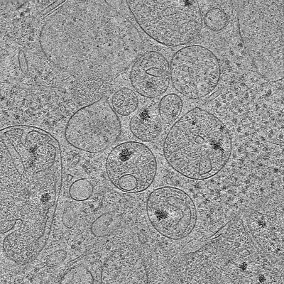 emd_4492.png emd_4492.png | 141.8 KB | ||
| Archive directory |  http://ftp.pdbj.org/pub/emdb/structures/EMD-4492ftp://ftp.pdbj.org/pub/emdb/structures/EMD-4492 http://ftp.pdbj.org/pub/emdb/structures/EMD-4492ftp://ftp.pdbj.org/pub/emdb/structures/EMD-4492 | HTTPS FTP |
-Related structure data

-Links
| EMDB pages | EMDB (EBI/PDBe) / EMDataResource |
|---|
-Map
| File | Download / File: emd_4492.map.gz / Format: CCP4 / Size: 2.2 GB / Type: IMAGE STORED AS SIGNED BYTE | ||||||||||||||||||||||||||||||||||||||||||||||||||||||||||||
|---|---|---|---|---|---|---|---|---|---|---|---|---|---|---|---|---|---|---|---|---|---|---|---|---|---|---|---|---|---|---|---|---|---|---|---|---|---|---|---|---|---|---|---|---|---|---|---|---|---|---|---|---|---|---|---|---|---|---|---|---|---|
| Annotation | Reconstructed electron cryo-tomogram of mitochondria in HeLa cells overexpression GFP-Bax | ||||||||||||||||||||||||||||||||||||||||||||||||||||||||||||
| Voxel size | X=Y=Z: 7.506 Å | ||||||||||||||||||||||||||||||||||||||||||||||||||||||||||||
| Density |
| ||||||||||||||||||||||||||||||||||||||||||||||||||||||||||||
| Symmetry | Space group: 1 | ||||||||||||||||||||||||||||||||||||||||||||||||||||||||||||
| Details | EMDB XML:
CCP4 map header:
| ||||||||||||||||||||||||||||||||||||||||||||||||||||||||||||
-Supplemental data
- Sample components
-Entire : Bax/Bak double knockout cell (homo sapiens) stably expressing GFP-Bax
| Entire | Name: Bax/Bak double knockout cell (homo sapiens) stably expressing GFP-Bax |
|---|---|
| Components |
|
-Supramolecule #1: Bax/Bak double knockout cell (homo sapiens) stably expressing GFP-Bax
| Supramolecule | Name: Bax/Bak double knockout cell (homo sapiens) stably expressing GFP-Bax type: cell / ID: 1 / Parent: 0 Details: Cryofixation of cell was performed 3 h after treatment with ABT-737 |
|---|---|
| Source (natural) | Organism: Homo sapiens (human) |
-Experimental details
-Structure determination
| Method | cryo EM |
|---|---|
 Processing Processing | electron tomography |
| Aggregation state | cell |
-Sample preparation
| Buffer | pH: 7.4 Details: DMEM, high glucose, GlutaMAX, pyruvate (Thermo 31996) medium supplemented with 10% heat-inactivated FBS (Gibco 10270), 10 mM HEPES, and 1x NEAA (Thermo 11140) |
|---|---|
| Grid | Model: Quantifoil R2/2 / Material: GOLD / Mesh: 200 / Support film - Material: CARBON / Support film - topology: HOLEY |
| Vitrification | Cryogen name: ETHANE / Instrument: HOMEMADE PLUNGER Details: Grids were manually backside blotted using Whatman filter paper No. 1 and vitrified using a manual plunger.. |
| Details | Bax/Bak DKO HCT116 GFP-Bax cells were grown on grids for 36 h, stained with MitoTracker Deep Red, and incubated with ABT-737 and Q-VD-OPh for 3 h before plunge-freezing. Grids were manually backside blotted using Whatman filter paper No. 1 and vitrified using a manual plunger. |
| Sectioning | Focused ion beam - Instrument: OTHER / Focused ion beam - Ion: OTHER / Focused ion beam - Voltage: 30 kV / Focused ion beam - Current: 1 nA / Focused ion beam - Duration: 10800 sec. / Focused ion beam - Temperature: 83 K / Focused ion beam - Initial thickness: 1000 nm / Focused ion beam - Final thickness: 200 nm Focused ion beam - Details: Cells were cryo-FIB milled to prepare lamellae using a Scios DualBeam FIB/SEM (FEI) equipped with a Quorum cryo-stage (PP3010T), following the protocol described in ...Focused ion beam - Details: Cells were cryo-FIB milled to prepare lamellae using a Scios DualBeam FIB/SEM (FEI) equipped with a Quorum cryo-stage (PP3010T), following the protocol described in Schaffer et al. (2015). In brief, grids were coated with an organic Pt compound using the gas injection system for either 8 s at 12 mm working distance or 30 s at 13 mm working distance from a stage tilt of 25 degrees. The stage was then tilted so that the grid was at a 10 degree angle towards the ion beam for all subsequent steps. The electron beam was used at 13 pA and 5-10 kV to locate cells, 2 kV for subsequent imaging. The ion beam was used at 30 kV and 10 pA for imaging. Rough milling was performed at 30 kV ion beam voltage, and subsequently the current was reduced from 0.5 nA to 0.3 nA until a lamella thickness of 5 um was reached, and further to 0.1 nA until 1 um lamella thickness. Fine milling to a final lamella thickness of approximately 200 nm was performed either at 30 kV and 30 pA, or 16 kV and 11 pA ion beam setting.. The value given for _emd_sectioning_focused_ion_beam.instrument is FEI Scios DualBeam FIB/SEM. This is not in a list of allowed values set(['DB235', 'OTHER']) so OTHER is written into the XML file. |
- Electron microscopy
| Microscope | FEI TITAN KRIOS |
|---|---|
| Specialist optics | Energy filter - Name: GIF Quantum LS / Energy filter - Slit width: 20 eV |
| Details | Montaged images of the entire grid were acquired at low magnification at pixel size of either 190.9 or 99.4 nm. Intermediate magnification maps of lamella were acquired at pixel size 5.5 nm. |
| Image recording | Film or detector model: GATAN K2 SUMMIT (4k x 4k) / Detector mode: COUNTING / Digitization - Dimensions - Width: 3710 pixel / Digitization - Dimensions - Height: 3838 pixel / Average electron dose: 1.1 e/Å2 Details: Electron cryo-tomographic tilt-series were collected on a Titan Krios (FEI) operated at 300 kV using a Quantum energy filter (slit width 20 eV) and a K2 direct electron detector (Gatan) in ...Details: Electron cryo-tomographic tilt-series were collected on a Titan Krios (FEI) operated at 300 kV using a Quantum energy filter (slit width 20 eV) and a K2 direct electron detector (Gatan) in counting mode at a pixel size of 3.7 angstroms and at a dose rate of ~ 2-4 e-/pixel/second on the detector, dependent on sample thickness. Tilt-series were acquired between +/- 60 degrees starting from 0 degrees with 1 degrees increment using SerialEM (Mastronarde, 2005) following a grouped dose-symmetric acquisition with a group size of 4 (Bharat et al., 2018; Hagen et al., 2017), and at -5 um defocus. A dose of approximately 1.0 to 1.2 e-/square angstroms was applied per image of the tilt-series. |
| Electron beam | Acceleration voltage: 300 kV / Electron source:  FIELD EMISSION GUN FIELD EMISSION GUN |
| Electron optics | Illumination mode: FLOOD BEAM / Imaging mode: BRIGHT FIELD / Nominal defocus max: 5.0 µm / Nominal defocus min: 5.0 µm |
| Sample stage | Specimen holder model: FEI TITAN KRIOS AUTOGRID HOLDER / Cooling holder cryogen: NITROGEN |
| Experimental equipment |  Model: Titan Krios / Image courtesy: FEI Company |
-Image processing
| Final reconstruction | Algorithm: SIMULTANEOUS ITERATIVE (SIRT) / Software - Name: eTomo (ver. 4.10.20) / Details: 10 iterations / Number images used: 99 |
|---|
