 ムービー
ムービー コントローラー


+ データを開く

- 基本情報
基本情報
| 登録情報 | データベース: EMDB / ID: EMD-3967 | |||||||||
|---|---|---|---|---|---|---|---|---|---|---|
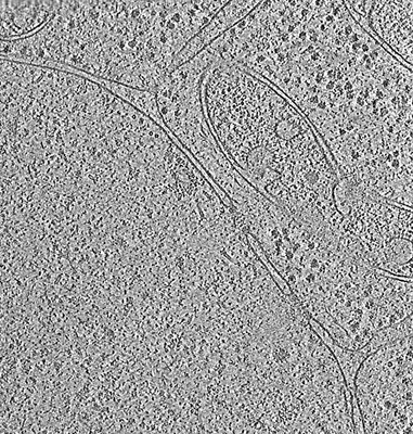
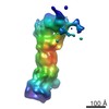
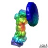
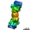
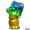
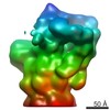
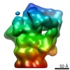
| タイトル | In situ cryo-electron tomogram from Chlamydomonas reinhardtii of the cellular environment around the nuclear envelope | |||||||||
 マップデータ マップデータ | Cryo-electron tomogram of Chlamydomonas reinhardtii nuclear membrane | |||||||||
 試料 試料 |
| |||||||||
| 生物種 |    Chlamydomonas reinhardtii (クラミドモナス) Chlamydomonas reinhardtii (クラミドモナス) | |||||||||
| 手法 | 電子線トモグラフィー法 / クライオ電子顕微鏡法 | |||||||||
 データ登録者 データ登録者 | Albert S / Schaffer M / Beck F / Mosalaganti S / Asano S / Thomas HF / Plitzko J / Beck M / Baumeister W / Engel BD | |||||||||
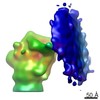
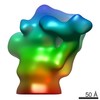
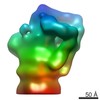
 引用 引用 | ジャーナル: Proc Natl Acad Sci U S A / 年: 2017 タイトル: Proteasomes tether to two distinct sites at the nuclear pore complex. 著者: Sahradha Albert / Miroslava Schaffer / Florian Beck / Shyamal Mosalaganti / Shoh Asano / Henry F Thomas / Jürgen M Plitzko / Martin Beck / Wolfgang Baumeister / Benjamin D Engel /  要旨: The partitioning of cellular components between the nucleus and cytoplasm is the defining feature of eukaryotic life. The nuclear pore complex (NPC) selectively gates the transport of macromolecules ...The partitioning of cellular components between the nucleus and cytoplasm is the defining feature of eukaryotic life. The nuclear pore complex (NPC) selectively gates the transport of macromolecules between these compartments, but it is unknown whether surveillance mechanisms exist to reinforce this function. By leveraging in situ cryo-electron tomography to image the native cellular environment of , we observed that nuclear 26S proteasomes crowd around NPCs. Through a combination of subtomogram averaging and nanometer-precision localization, we identified two classes of proteasomes tethered via their Rpn9 subunits to two specific NPC locations: binding sites on the NPC basket that reflect its eightfold symmetry and more abundant binding sites at the inner nuclear membrane that encircle the NPC. These basket-tethered and membrane-tethered proteasomes, which have similar substrate-processing state frequencies as proteasomes elsewhere in the cell, are ideally positioned to regulate transcription and perform quality control of both soluble and membrane proteins transiting the NPC. | |||||||||
| 履歴 |
|
- 構造の表示
構造の表示
| ムービー |
 ムービービューア ムービービューア |
|---|---|
| 構造ビューア | EMマップ: SurfViewMolmilJmol/JSmol |
| 添付画像 |
- ダウンロードとリンク
-EMDBアーカイブ
| マップデータ | emd_3967.map.gz | 340 MB | EMDBマップデータ形式 | |
|---|---|---|---|---|
| ヘッダ (付随情報) | emd-3967-v30.xmlemd-3967.xml | 14.3 KB 14.3 KB | 表示 表示 | EMDBヘッダ |
| 画像 |  emd_3967.png emd_3967.png | 164.6 KB | ||
| アーカイブディレクトリ |  http://ftp.pdbj.org/pub/emdb/structures/EMD-3967ftp://ftp.pdbj.org/pub/emdb/structures/EMD-3967 http://ftp.pdbj.org/pub/emdb/structures/EMD-3967ftp://ftp.pdbj.org/pub/emdb/structures/EMD-3967 | HTTPS FTP |
-関連構造データ
-リンク
| EMDBのページ | EMDB (EBI/PDBe) / EMDataResource |
|---|
-マップ
| ファイル | ダウンロード / ファイル: emd_3967.map.gz / 形式: CCP4 / 大きさ: 762.2 MB / タイプ: IMAGE STORED AS SIGNED INTEGER (2 BYTES) | ||||||||||||||||||||||||||||||||||||||||||||||||||||||||||||
|---|---|---|---|---|---|---|---|---|---|---|---|---|---|---|---|---|---|---|---|---|---|---|---|---|---|---|---|---|---|---|---|---|---|---|---|---|---|---|---|---|---|---|---|---|---|---|---|---|---|---|---|---|---|---|---|---|---|---|---|---|---|
| 注釈 | Cryo-electron tomogram of Chlamydomonas reinhardtii nuclear membrane | ||||||||||||||||||||||||||||||||||||||||||||||||||||||||||||
| ボクセルのサイズ | X=Y=Z: 13.68 Å | ||||||||||||||||||||||||||||||||||||||||||||||||||||||||||||
| 密度 |
| ||||||||||||||||||||||||||||||||||||||||||||||||||||||||||||
| 対称性 | 空間群: 1 | ||||||||||||||||||||||||||||||||||||||||||||||||||||||||||||
| 詳細 | EMDB XML:
CCP4マップ ヘッダ情報:
| ||||||||||||||||||||||||||||||||||||||||||||||||||||||||||||
-添付データ
- 試料の構成要素
-全体 : Whole Chlamydomonas cells
| 全体 | 名称: Whole Chlamydomonas cells |
|---|---|
| 要素 |
|
-超分子 #1: Whole Chlamydomonas cells
| 超分子 | 名称: Whole Chlamydomonas cells / タイプ: cell / ID: 1 / 親要素: 0 詳細: Grown suspended in TAP media, with normal atmosphere aeration and constant light |
|---|---|
| 由来(天然) | 生物種: Chlamydomonas reinhardtii (クラミドモナス) 株: mat3-4 |
-実験情報
-構造解析
| 手法 | クライオ電子顕微鏡法 |
|---|---|
 解析 解析 | 電子線トモグラフィー法 |
| 試料の集合状態 | cell |
-試料調製
| 緩衝液 | pH: 7 / 詳細: TAP media |
|---|---|
| グリッド | モデル: Quantifoil R2/1 / 材質: COPPER / メッシュ: 200 / 前処理 - タイプ: GLOW DISCHARGE / 前処理 - 雰囲気: AIR |
| 凍結 | 凍結剤: ETHANE-PROPANE / チャンバー内湿度: 90 % / チャンバー内温度: 293 K / 装置: FEI VITROBOT MARK IV 詳細: Blotted for 10 seconds with 10 blot force before plunging.. |
| 詳細 | The cells were frozen onto grids, then thinned using cryo-focused ion beam milling. |
| 切片作成 | 集束イオンビーム - 装置: OTHER / 集束イオンビーム - イオン: OTHER / 集束イオンビーム - 電圧: 30 kV / 集束イオンビーム - 電流: 0.03 nA / 集束イオンビーム - Dose rate: 2 / 集束イオンビーム - 時間: 2400 sec. / 集束イオンビーム - 温度: 80 K / 集束イオンビーム - Initial thickness: 5000 / 集束イオンビーム - 最終 厚さ: 100 集束イオンビーム - 詳細: Starting curent: 0.5 nA, Final current: 0.03 nA. The value given for _emd_sectioning_focused_ion_beam.instrument is FEI Scios DB-FIB. This is not in a list of ...集束イオンビーム - 詳細: Starting curent: 0.5 nA, Final current: 0.03 nA. The value given for _emd_sectioning_focused_ion_beam.instrument is FEI Scios DB-FIB. This is not in a list of allowed values set(['DB235', 'OTHER']) so OTHER is written into the XML file. |
- 電子顕微鏡法
| 顕微鏡 | FEI TITAN KRIOS |
|---|---|
| 電子線 | 加速電圧: 300 kV / 電子線源: FIELD EMISSION GUN |
| 電子光学系 | C2レンズ絞り径: 70.0 µm / 照射モード: FLOOD BEAM / 撮影モード: BRIGHT FIELDBright-field microscopy / Cs: 2.7 mm / 最大 デフォーカス(公称値): 6.0 µm / 最小 デフォーカス(公称値): 4.0 µm / 倍率(公称値): 42000 |
| 特殊光学系 | エネルギーフィルター - 名称: GIF Quantum LS エネルギーフィルター - エネルギー下限: 0 eV エネルギーフィルター - エネルギー上限: 20 eV |
| 試料ステージ | 試料ホルダーモデル: FEI TITAN KRIOS AUTOGRID HOLDER ホルダー冷却材: NITROGEN |
| 撮影 | フィルム・検出器のモデル: GATAN K2 SUMMIT (4k x 4k) 検出モード: COUNTING / デジタル化 - サイズ - 横: 3838 pixel / デジタル化 - サイズ - 縦: 3710 pixel / 平均露光時間: 1.5 sec. / 平均電子線量: 1.5 e/Å2 詳細: Images were collected in movie mode at 12 frames per second |
| 実験機器 |  モデル: Titan Krios / 画像提供: FEI Company |
-画像解析
| CTF補正 | ソフトウェア - 名称: IMOD |
|---|---|
| 最終 再構成 | アルゴリズム: BACK PROJECTION / ソフトウェア - 名称: IMOD / 詳細: This is a bin4 (twice binned) tomogram. / 使用した粒子像数: 61 |
