 Movie
Movie Controller
 Structure viewers
 About Yorodumi Papers


+Search query
-Structure paper
| Title | Proteasomes tether to two distinct sites at the nuclear pore complex. |
|---|---|
| Journal, issue, pages | Proc Natl Acad Sci U S A, Vol. 114, Issue 52, Page 13726-13731, Year 2017 |
| Publish date | Dec 26, 2017 |
 Authors Authors | Sahradha Albert / Miroslava Schaffer / Florian Beck / Shyamal Mosalaganti / Shoh Asano / Henry F Thomas / Jürgen M Plitzko / Martin Beck / Wolfgang Baumeister / Benjamin D Engel /  |
| PubMed Abstract | The partitioning of cellular components between the nucleus and cytoplasm is the defining feature of eukaryotic life. The nuclear pore complex (NPC) selectively gates the transport of macromolecules ...The partitioning of cellular components between the nucleus and cytoplasm is the defining feature of eukaryotic life. The nuclear pore complex (NPC) selectively gates the transport of macromolecules between these compartments, but it is unknown whether surveillance mechanisms exist to reinforce this function. By leveraging in situ cryo-electron tomography to image the native cellular environment of , we observed that nuclear 26S proteasomes crowd around NPCs. Through a combination of subtomogram averaging and nanometer-precision localization, we identified two classes of proteasomes tethered via their Rpn9 subunits to two specific NPC locations: binding sites on the NPC basket that reflect its eightfold symmetry and more abundant binding sites at the inner nuclear membrane that encircle the NPC. These basket-tethered and membrane-tethered proteasomes, which have similar substrate-processing state frequencies as proteasomes elsewhere in the cell, are ideally positioned to regulate transcription and perform quality control of both soluble and membrane proteins transiting the NPC. |
 External links External links | Proc Natl Acad Sci U S A / PubMed:29229809 / PubMed Central |
| Methods | EM (subtomogram averaging) / EM (tomography) |
| Resolution | 23.81 - 36.48 Å |
| Structure data | 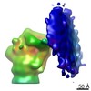 EMDB-3936: 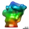 EMDB-3937: 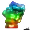 EMDB-3938: 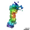 EMDB-3939: 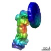 EMDB-3940:  EMDB-3967: |
| Source |
|

 Chlamydomonas reinhardtii (plant)