 ムービー
ムービー コントローラー


+ データを開く

- 基本情報
基本情報
| 登録情報 | データベース: EMDB / ID: EMD-3804 | |||||||||
|---|---|---|---|---|---|---|---|---|---|---|
| タイトル | Cryo-EM structure of the yeast chromatin modifying complex SAGA | |||||||||
 マップデータ マップデータ | ||||||||||
 試料 試料 |
| |||||||||
| 生物種 |   Komagataella pastoris (菌類) Komagataella pastoris (菌類) | |||||||||
| 手法 | 単粒子再構成法 / クライオ電子顕微鏡法 / 解像度: 11.7 Å | |||||||||
 データ登録者 データ登録者 | Sharov G / Voltz K / Durand A / Kolesnikova O / Dejaegere A / Papai G / Myasnikov AG / Ben-Shem A / Schultz P | |||||||||
 引用 引用 | ジャーナル: Nat Commun / 年: 2017 タイトル: Structure of the transcription activator target Tra1 within the chromatin modifying complex SAGA. 著者: Grigory Sharov / Karine Voltz / Alexandre Durand / Olga Kolesnikova / Gabor Papai / Alexander G Myasnikov / Annick Dejaegere / Adam Ben Shem / Patrick Schultz /    要旨: The transcription co-activator complex SAGA is recruited to gene promoters by sequence-specific transcriptional activators and by chromatin modifications to promote pre-initiation complex formation. ...The transcription co-activator complex SAGA is recruited to gene promoters by sequence-specific transcriptional activators and by chromatin modifications to promote pre-initiation complex formation. The yeast Tra1 subunit is the major target of acidic activators such as Gal4, VP16, or Gcn4 but little is known about its structural organization. The 430 kDa Tra1 subunit and its human homolog the transformation/transcription domain-associated protein TRRAP are members of the phosphatidyl 3-kinase-related kinase (PIKK) family. Here, we present the cryo-EM structure of the entire SAGA complex where the major target of activator binding, the 430 kDa Tra1 protein, is resolved with an average resolution of 5.7 Å. The high content of alpha-helices in Tra1 enabled tracing of the majority of its main chain. Our results highlight the integration of Tra1 within the major epigenetic regulator SAGA. | |||||||||
| 履歴 |
|
- 構造の表示
構造の表示
| ムービー |
 ムービービューア ムービービューア |
|---|---|
| 構造ビューア | EMマップ: SurfViewMolmilJmol/JSmol |
| 添付画像 |

- ダウンロードとリンク
-EMDBアーカイブ
| マップデータ | emd_3804.map.gz | 410.8 MB | EMDBマップデータ形式 | |
|---|---|---|---|---|
| ヘッダ (付随情報) | emd-3804-v30.xmlemd-3804.xml | 13.7 KB 13.7 KB | 表示 表示 | EMDBヘッダ |
| FSC (解像度算出) | emd_3804_fsc.xml | 17.9 KB | 表示 | FSCデータファイル |
| 画像 | 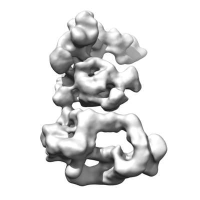 emd_3804.png emd_3804.png | 48 KB | ||
| アーカイブディレクトリ |  http://ftp.pdbj.org/pub/emdb/structures/EMD-3804ftp://ftp.pdbj.org/pub/emdb/structures/EMD-3804 http://ftp.pdbj.org/pub/emdb/structures/EMD-3804ftp://ftp.pdbj.org/pub/emdb/structures/EMD-3804 | HTTPS FTP |
-関連構造データ


-リンク
| EMDBのページ | EMDB (EBI/PDBe) / EMDataResource |
|---|
-マップ
| ファイル | ダウンロード / ファイル: emd_3804.map.gz / 形式: CCP4 / 大きさ: 512 MB / タイプ: IMAGE STORED AS FLOATING POINT NUMBER (4 BYTES) | ||||||||||||||||||||||||||||||||||||||||||||||||||||||||||||
|---|---|---|---|---|---|---|---|---|---|---|---|---|---|---|---|---|---|---|---|---|---|---|---|---|---|---|---|---|---|---|---|---|---|---|---|---|---|---|---|---|---|---|---|---|---|---|---|---|---|---|---|---|---|---|---|---|---|---|---|---|---|
| ボクセルのサイズ | X=Y=Z: 1.1 Å | ||||||||||||||||||||||||||||||||||||||||||||||||||||||||||||
| 密度 |
| ||||||||||||||||||||||||||||||||||||||||||||||||||||||||||||
| 対称性 | 空間群: 1 | ||||||||||||||||||||||||||||||||||||||||||||||||||||||||||||
| 詳細 | EMDB XML:
CCP4マップ ヘッダ情報:
| ||||||||||||||||||||||||||||||||||||||||||||||||||||||||||||
-添付データ
- 試料の構成要素
-全体 : Spt-Ada-Gcn5-acetyltransferase complex (SAGA)
| 全体 | 名称: Spt-Ada-Gcn5-acetyltransferase complex (SAGA) |
|---|---|
| 要素 |
|
-超分子 #1: Spt-Ada-Gcn5-acetyltransferase complex (SAGA)
| 超分子 | 名称: Spt-Ada-Gcn5-acetyltransferase complex (SAGA) / タイプ: complex / ID: 1 / 親要素: 0 / 含まれる分子: all |
|---|---|
| 由来(天然) | 生物種: Komagataella pastoris (菌類) |
| 分子量 | 理論値: 1.8 MDa |
-分子 #1: Cryo-EM structure of yeast SAGA complex
| 分子 | 名称: Cryo-EM structure of yeast SAGA complex / タイプ: protein_or_peptide / ID: 1 / 光学異性体: LEVO |
|---|---|
| 由来(天然) | 生物種: Komagataella pastoris (菌類) |
| 配列 | 文字列: () |
-実験情報
-構造解析
| 手法 | クライオ電子顕微鏡法 |
|---|---|
 解析 解析 | 単粒子再構成法 |
| 試料の集合状態 | particle |
-試料調製
| 濃度 | 0.2 mg/mL |
|---|---|
| 緩衝液 | pH: 8 |
| グリッド | モデル: Quantifoil R2/2 / 材質: COPPER / メッシュ: 300 / 支持フィルム - 材質: CARBON / 支持フィルム - トポロジー: HOLEY / 前処理 - タイプ: GLOW DISCHARGE / 前処理 - 雰囲気: AIR / 前処理 - 気圧: 0.018 kPa |
| 凍結 | 凍結剤: ETHANE / チャンバー内湿度: 95 % / チャンバー内温度: 277 K / 装置: FEI VITROBOT MARK IV / 詳細: Blot for 1 second before plunging. |
- 電子顕微鏡法
| 顕微鏡 | FEI TITAN KRIOS |
|---|---|
| 電子線 | 加速電圧: 300 kV / 電子線源: FIELD EMISSION GUN |
| 電子光学系 | C2レンズ絞り径: 100.0 µm / 最大 デフォーカス(補正後): 3.4 µm / 最小 デフォーカス(補正後): 1.4 µm / 倍率(補正後): 127272 / 照射モード: FLOOD BEAM / 撮影モード: BRIGHT FIELDBright-field microscopy / Cs: 0.001 mm / 最大 デフォーカス(公称値): 3.4 µm / 最小 デフォーカス(公称値): 1.4 µm / 倍率(公称値): 59000 |
| 特殊光学系 | 球面収差補正装置: Microscope has a Cs corrector |
| 試料ステージ | 試料ホルダーモデル: FEI TITAN KRIOS AUTOGRID HOLDER ホルダー冷却材: NITROGEN |
| 温度 | 最低: 70.0 K / 最高: 80.0 K |
| 撮影 | フィルム・検出器のモデル: FEI FALCON II (4k x 4k) 検出モード: INTEGRATING / デジタル化 - サイズ - 横: 4096 pixel / デジタル化 - サイズ - 縦: 4096 pixel / デジタル化 - サンプリング間隔: 14.0 µm / デジタル化 - 画像ごとのフレーム数: 2-8 / 撮影したグリッド数: 4 / 実像数: 8505 / 平均露光時間: 1.0 sec. / 平均電子線量: 60.0 e/Å2 詳細: Images were collected in movie-mode at 17 frames per second, frame 1 was not acquired. Every two frames were joined together, producing 8 frames per second. |
| 実験機器 |  モデル: Titan Krios / 画像提供: FEI Company |
-画像解析
| 粒子像選択 | 選択した数: 264901 | ||||||
|---|---|---|---|---|---|---|---|
| CTF補正 | ソフトウェア:
詳細: Full CTF correction in Relion | ||||||
| 初期モデル | モデルのタイプ: RANDOM CONICAL TILT 詳細: RCT model from previous study: DOI: 10.1016/j.molcel.2004.06.005 | ||||||
| 初期 角度割当 | タイプ: NOT APPLICABLE | ||||||
| 最終 角度割当 | タイプ: PROJECTION MATCHING Projection matching processing - Merit function: Maximum likelihood (ML ソフトウェア - 名称: RELION (ver. 1.4) | ||||||
| 最終 再構成 | 想定した対称性 - 点群: C1 (非対称) / アルゴリズム: FOURIER SPACE / 解像度のタイプ: BY AUTHOR / 解像度: 11.7 Å / 解像度の算出法: FSC 0.143 CUT-OFF / ソフトウェア - 名称: RELION (ver. 1.4) / 使用した粒子像数: 105916 | ||||||
| FSC曲線 (解像度の算出) | 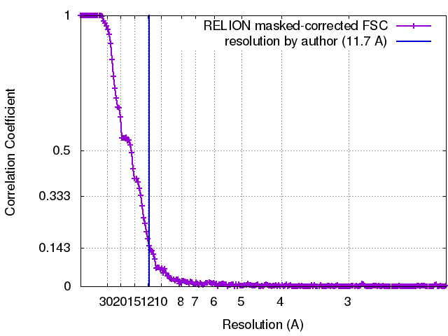 |
