National Institutes of Health/National Institute of General Medical Sciences
GM118099
米国
National Institutes of Health/National Institute of General Medical Sciences
GM118106
米国
引用
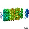
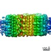
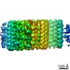
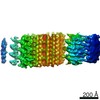
ジャーナル: J Cell Biol / 年: 2020 タイトル: Electron cryotomography of intact motile cilia defines the basal body to axoneme transition. 著者: Garrett A Greenan / Ronald D Vale / David A Agard / 要旨: Cells use motile cilia to generate force in the extracellular space. The structure of a cilium can be classified into three subdomains: the intracellular basal body (BB) that templates cilium ...Cells use motile cilia to generate force in the extracellular space. The structure of a cilium can be classified into three subdomains: the intracellular basal body (BB) that templates cilium formation, the extracellular axoneme that generates force, and the transition zone (TZ) that bridges them. While the BB is composed of triplet microtubules (TMTs), the axoneme is composed of doublet microtubules (DMTs), meaning the cilium must convert between different microtubule geometries. Here, we performed electron cryotomography to define this conversion, and our reconstructions reveal identifying structural features of the BB, TZ, and axoneme. Each region is distinct in terms of microtubule number and geometry, microtubule inner proteins, and microtubule linkers. TMT to DMT conversion occurs within the BB, and microtubule geometry changes to axonemal by the end of the TZ, followed by the addition of axoneme-specific components essential for cilium motility. Our results provide the highest-resolution images of the motile cilium to date and reveal how BBs template axonemes.
 ムービー
ムービー コントローラー


 データを開く
 基本情報
基本情報 マップデータ
 試料
試料
 データ登録者
 米国, 2件
  引用
引用 構造の表示
構造の表示 ムービービューア
 ダウンロードとリンク
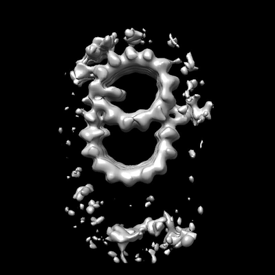 emd_20684.png
 http://ftp.pdbj.org/pub/emdb/structures/EMD-20684

 試料の構成要素
 解析
解析 電子顕微鏡法
 FIELD EMISSION GUN
