positive regulation of rRNA processing / 核様体 / small ribosomal subunit rRNA binding / ribosomal small subunit assembly / rRNA processing / cytosolic small ribosomal subunit / large ribosomal subunit rRNA binding / large ribosomal subunit / small ribosomal subunit / cytoplasmic translation ...positive regulation of rRNA processing / 核様体 / small ribosomal subunit rRNA binding / ribosomal small subunit assembly / rRNA processing / cytosolic small ribosomal subunit / large ribosomal subunit rRNA binding / large ribosomal subunit / small ribosomal subunit / cytoplasmic translation / 5S rRNA binding / cytosolic large ribosomal subunit / transferase activity / tRNA binding / negative regulation of translation / rRNA binding / リボソーム / structural constituent of ribosome / 翻訳 (生物学) / ribonucleoprotein complex / response to antibiotic / mRNA binding / DNA binding / RNA binding / zinc ion binding / metal ion binding / 細胞質基質 / 細胞質 類似検索 - 分子機能
Ribosomal protein S14, type Z / Ribosomal protein L31 type A / Ribosomal protein L31 signature. / Ribosomal protein L31 / Ribosomal protein L31 superfamily / Ribosomal protein L31 / Ribosomal protein L21, conserved site / Ribosomal protein L21 signature. / Ribosomal protein L16 signature 1. / : ...Ribosomal protein S14, type Z / Ribosomal protein L31 type A / Ribosomal protein L31 signature. / Ribosomal protein L31 / Ribosomal protein L31 superfamily / Ribosomal protein L31 / Ribosomal protein L21, conserved site / Ribosomal protein L21 signature. / Ribosomal protein L16 signature 1. / : / Ribosomal protein L6, conserved site / Ribosomal protein L6 signature 1. / Ribosomal protein L16, conserved site / Ribosomal protein L16 signature 2. / Ribosomal protein L17 signature. / Ribosomal protein S14/S29 / Ribosomal protein L28/L24 superfamily / Ribosomal protein L36 signature. / Ribosomal protein L32p, bacterial type / Ribosomal protein L28 / Ribosomal protein L35, conserved site / Ribosomal protein L35 signature. / Ribosomal protein L33, conserved site / Ribosomal protein L33 signature. / Ribosomal protein L35, non-mitochondrial / Ribosomal protein L5, bacterial-type / Ribosomal protein L6, bacterial-type / Ribosomal protein L18, bacterial-type / Ribosomal protein L19, conserved site / Ribosomal protein L19 signature. / Ribosomal protein L36 / Ribosomal protein L36 superfamily / Ribosomal protein L36 / Ribosomal protein L20 signature. / Ribosomal protein S3, bacterial-type / Ribosomal protein S6, conserved site / Ribosomal protein S6 signature. / Ribosomal protein L27, conserved site / Ribosomal protein L27 signature. / Ribosomal protein S19, bacterial-type / Ribosomal protein S7, bacterial/organellar-type / Ribosomal protein S11, bacterial-type / Ribosomal protein S13, bacterial-type / Ribosomal protein S20 / Ribosomal protein S20 superfamily / Ribosomal protein S20 / Ribosomal protein S9, bacterial/plastid / Ribosomal protein L14P, bacterial-type / Ribosomal protein L34, conserved site / Ribosomal protein L34 signature. / 30S ribosomal protein S17 / Ribosomal protein S5, bacterial-type / Ribosomal protein L22, bacterial/chloroplast-type / Ribosomal protein S6, plastid/chloroplast / Ribosomal protein L35 / Ribosomal protein L35 superfamily / Ribosomal protein L2, bacterial/organellar-type / Ribosomal protein L35 / Ribosomal L28 family / Ribosomal protein L33 / Ribosomal protein L33 / Ribosomal protein L28/L24 / Ribosomal protein L33 superfamily / : / Ribosomal protein L30, bacterial-type / Ribosomal protein L16 / Ribosomal protein L18 / Ribosomal L18 of archaea, bacteria, mitoch. and chloroplast / Ribosomal protein S18, conserved site / Ribosomal protein S18 signature. / L28p-like / Ribosomal protein L20 / Ribosomal protein L20 / Ribosomal protein L20, C-terminal / Ribosomal protein L21 / Ribosomal protein L27 / Ribosomal L27 protein / Ribosomal protein L19 / Ribosomal protein L19 superfamily / Ribosomal protein L19 / Ribosomal protein S15, bacterial-type / Ribosomal proteins 50S L24/mitochondrial 39S L24 / Ribosomal protein L17 / Ribosomal protein L17 superfamily / Ribosomal protein L17 / Ribosomal protein L21-like / L21-like superfamily / Ribosomal prokaryotic L21 protein / Ribosomal L32p protein family / Ribosomal protein S6 / Ribosomal protein S6 / Ribosomal protein S6 superfamily / Ribosomal protein L24 / Ribosomal protein L32p / Ribosomal protein S12, bacterial-type / Ribosomal protein L34 / Ribosomal protein L34 / Ribosomal protein L13, bacterial-type / Translation elongation factor EF1B/ribosomal protein S6 / Ribosomal protein L23/L25, conserved site 類似検索 - ドメイン・相同性
Large ribosomal subunit protein bL19 / Large ribosomal subunit protein bL32 / Small ribosomal subunit protein uS11 / Large ribosomal subunit protein bL34 / Large ribosomal subunit protein bL27 / Large ribosomal subunit protein uL24 / Large ribosomal subunit protein uL29 / Small ribosomal subunit protein uS17 / Large ribosomal subunit protein uL14 / Large ribosomal subunit protein uL5 ...Large ribosomal subunit protein bL19 / Large ribosomal subunit protein bL32 / Small ribosomal subunit protein uS11 / Large ribosomal subunit protein bL34 / Large ribosomal subunit protein bL27 / Large ribosomal subunit protein uL24 / Large ribosomal subunit protein uL29 / Small ribosomal subunit protein uS17 / Large ribosomal subunit protein uL14 / Large ribosomal subunit protein uL5 / Small ribosomal subunit protein uS14B / Small ribosomal subunit protein uS8 / Large ribosomal subunit protein uL16 / Large ribosomal subunit protein uL15 / Large ribosomal subunit protein uL30 / Large ribosomal subunit protein bL17 / Large ribosomal subunit protein bL36 / Small ribosomal subunit protein uS13 / Small ribosomal subunit protein uS3 / Small ribosomal subunit protein uS5 / Small ribosomal subunit protein bS6 / Small ribosomal subunit protein uS7 / Small ribosomal subunit protein uS9 / Small ribosomal subunit protein uS10 / Small ribosomal subunit protein uS12 / Small ribosomal subunit protein uS15 / Small ribosomal subunit protein bS18 / Small ribosomal subunit protein uS19 / Small ribosomal subunit protein bS20 / Large ribosomal subunit protein bL21 / Large ribosomal subunit protein bL28 / Large ribosomal subunit protein uL22 / Large ribosomal subunit protein uL2 / Large ribosomal subunit protein uL3 / Large ribosomal subunit protein uL4 / Large ribosomal subunit protein uL23 / Large ribosomal subunit protein uL6 / Large ribosomal subunit protein uL18 / Large ribosomal subunit protein bL20 / Large ribosomal subunit protein bL35 / Large ribosomal subunit protein bL33A / Large ribosomal subunit protein uL13 / Large ribosomal subunit protein bL31 類似検索 - 構成要素
ジャーナル: Nat Commun / 年: 2024 タイトル: RAPP-containing arrest peptides induce translational stalling by short circuiting the ribosomal peptidyltransferase activity. 著者: Martino Morici / Sara Gabrielli / Keigo Fujiwara / Helge Paternoga / Bertrand Beckert / Lars V Bock / Shinobu Chiba / Daniel N Wilson / 要旨: Arrest peptides containing RAPP (ArgAlaProPro) motifs have been discovered in both Gram-positive and Gram-negative bacteria, where they are thought to regulate expression of important protein ...Arrest peptides containing RAPP (ArgAlaProPro) motifs have been discovered in both Gram-positive and Gram-negative bacteria, where they are thought to regulate expression of important protein localization machinery components. Here we determine cryo-EM structures of ribosomes stalled on RAPP arrest motifs in both Bacillus subtilis and Escherichia coli. Together with molecular dynamics simulations, our structures reveal that the RAPP motifs allow full accommodation of the A-site tRNA, but prevent the subsequent peptide bond from forming. Our data support a model where the RAP in the P-site interacts and stabilizes a single hydrogen atom on the Pro-tRNA in the A-site, thereby preventing an optimal geometry for the nucleophilic attack required for peptide bond formation to occur. This mechanism to short circuit the ribosomal peptidyltransferase activity is likely to operate for the majority of other RAPP-like arrest peptides found across diverse bacterial phylogenies.
フィルム・検出器のモデル: GATAN K3 BIOQUANTUM (6k x 4k) 平均電子線量: 75.6 e/Å2
実験機器
モデル: Titan Krios / 画像提供: FEI Company
-
画像解析
初期モデル
モデルのタイプ: INSILICO MODEL In silico モデル: An unpublished map from our group was initially low-pass filtered and used as model, since the biology was analogous
初期 角度割当
タイプ: MAXIMUM LIKELIHOOD
最終 角度割当
タイプ: MAXIMUM LIKELIHOOD
最終 再構成
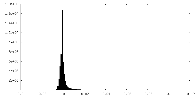
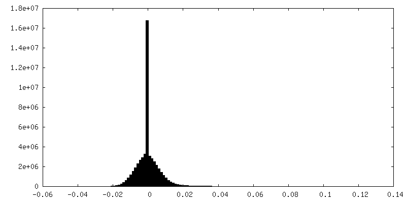
解像度のタイプ: BY AUTHOR / 解像度: 2.3 Å / 解像度の算出法: FSC 0.143 CUT-OFF / 使用した粒子像数: 142978
 ムービー
ムービー コントローラー


 データを開く
 基本情報
基本情報
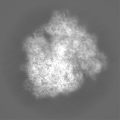
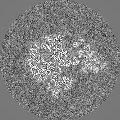
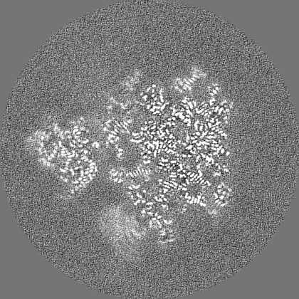
 マップデータ
 試料
試料 キーワード
キーワード Stalling / Nascent chain / elongation arrest /
  機能・相同性情報
 Amycolatopsis japonica (バクテリア) /
  データ登録者
 ドイツ, 1件
  引用
引用
 構造の表示
構造の表示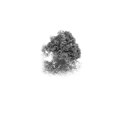
 ダウンロードとリンク
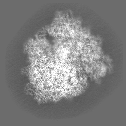
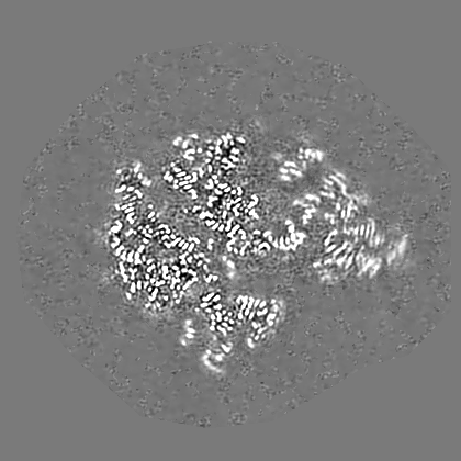
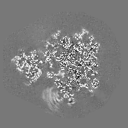
 emd_18332.png
 http://ftp.pdbj.org/pub/emdb/structures/EMD-18332


 Z
Z Y
Y X
X


 試料の構成要素

 解析
解析 電子顕微鏡法
