Phosphorylation of proteins involved in the G2/M transition by Cyclin A:Cdc2 complexes / cyclin A2-CDK1 complex / cell cycle G1/S phase transition / cellular response to luteinizing hormone stimulus / mitotic cell cycle phase transition / Transcription of E2F targets under negative control by p107 (RBL1) and p130 (RBL2) in complex with HDAC1 / cyclin-dependent protein serine/threonine kinase inhibitor activity / cellular response to leptin stimulus / male pronucleus / female pronucleus ...Phosphorylation of proteins involved in the G2/M transition by Cyclin A:Cdc2 complexes / cyclin A2-CDK1 complex / cell cycle G1/S phase transition / cellular response to luteinizing hormone stimulus / mitotic cell cycle phase transition / Transcription of E2F targets under negative control by p107 (RBL1) and p130 (RBL2) in complex with HDAC1 / cyclin-dependent protein serine/threonine kinase inhibitor activity / cellular response to leptin stimulus / male pronucleus / female pronucleus / cellular response to cocaine / response to glucagon / cyclin-dependent protein serine/threonine kinase regulator activity / cellular response to insulin-like growth factor stimulus / positive regulation of DNA biosynthetic process / cochlea development / cyclin A1-CDK2 complex / cyclin E2-CDK2 complex / cyclin E1-CDK2 complex / cellular response to platelet-derived growth factor stimulus / cyclin A2-CDK2 complex / positive regulation of DNA-templated DNA replication initiation / G2期 / cyclin-dependent protein kinase activity / Y染色体 / Phosphorylation of proteins involved in G1/S transition by active Cyclin E:Cdk2 complexes / positive regulation of heterochromatin formation / p53-Dependent G1 DNA Damage Response / X染色体 / PTK6 Regulates Cell Cycle / regulation of anaphase-promoting complex-dependent catabolic process / Defective binding of RB1 mutants to E2F1,(E2F2, E2F3) / regulation of DNA replication / centriole replication / Regulation of APC/C activators between G1/S and early anaphase / centrosome duplication / Telomere Extension By Telomerase / G0 and Early G1 / Activation of the pre-replicative complex / cyclin-dependent protein kinase holoenzyme complex / cellular response to nitric oxide / サイクリン依存性キナーゼ / animal organ regeneration / cyclin-dependent protein serine/threonine kinase activity / TP53 Regulates Transcription of Genes Involved in G1 Cell Cycle Arrest / カハール体 / Activation of ATR in response to replication stress / Cyclin E associated events during G1/S transition / Cyclin A/B1/B2 associated events during G2/M transition / Cyclin A:Cdk2-associated events at S phase entry / condensed chromosome / mitotic G1 DNA damage checkpoint signaling / regulation of mitotic cell cycle / regulation of G2/M transition of mitotic cell cycle / cyclin binding / post-translational protein modification / meiotic cell cycle / positive regulation of DNA replication / response to organic substance / male germ cell nucleus / cellular response to estradiol stimulus / Cdc20:Phospho-APC/C mediated degradation of Cyclin A / G1/S transition of mitotic cell cycle / potassium ion transport / DNA Damage/Telomere Stress Induced Senescence / CDK-mediated phosphorylation and removal of Cdc6 / SCF(Skp2)-mediated degradation of p27/p21 / 遺伝的組換え / Orc1 removal from chromatin / Transcriptional regulation of granulopoiesis / Cyclin D associated events in G1 / G2/M transition of mitotic cell cycle / positive regulation of fibroblast proliferation / 細胞老化 / Regulation of TP53 Degradation / 核膜 / Factors involved in megakaryocyte development and platelet production / Processing of DNA double-strand break ends / cellular response to hypoxia / Senescence-Associated Secretory Phenotype (SASP) / 遺伝子発現の調節 / peptidyl-serine phosphorylation / Ras protein signal transduction / Regulation of TP53 Activity through Phosphorylation / transcription regulator complex / DNA複製 / chromosome, telomeric region / regulation of cell cycle / Ub-specific processing proteases / エンドソーム / クロマチンリモデリング / protein domain specific binding / 細胞分裂 / protein phosphorylation / protein serine kinase activity / DNA修復 / protein serine/threonine kinase activity / 中心体 / DNA-templated transcription / positive regulation of cell population proliferation 類似検索 - 分子機能
Cyclin-dependent kinase inhibitor domain / Cyclin-dependent kinase inhibitor domain superfamily / サイクリン依存性キナーゼ阻害因子 / Cyclin-A, N-terminal APC/C binding region / Cyclin-A N-terminal APC/C binding region / : / Cyclin, C-terminal domain / : / Cyclins signature. / サイクリン ...Cyclin-dependent kinase inhibitor domain / Cyclin-dependent kinase inhibitor domain superfamily / サイクリン依存性キナーゼ阻害因子 / Cyclin-A, N-terminal APC/C binding region / Cyclin-A N-terminal APC/C binding region / : / Cyclin, C-terminal domain / : / Cyclins signature. / サイクリン / Cyclin, C-terminal domain / Cyclin_C / Cyclin, N-terminal / Cyclin, N-terminal domain / Cyclin-like / domain present in cyclins, TFIIB and Retinoblastoma / Cyclin-like superfamily / Serine/threonine-protein kinase, active site / Serine/Threonine protein kinases active-site signature. / Protein kinase domain / Serine/Threonine protein kinases, catalytic domain / Protein kinase, ATP binding site / Protein kinases ATP-binding region signature. / Protein kinase domain profile. / Protein kinase domain / Protein kinase-like domain superfamily 類似検索 - ドメイン・相同性
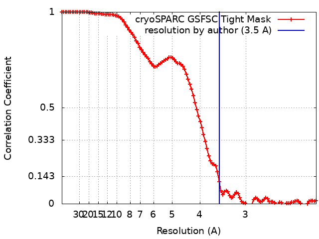
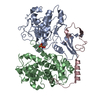
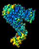
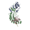
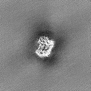
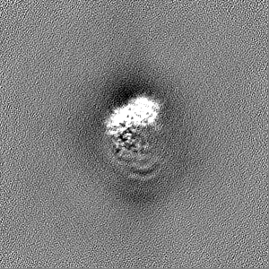
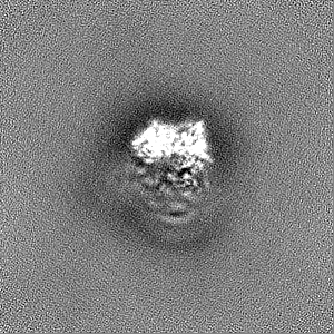
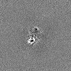
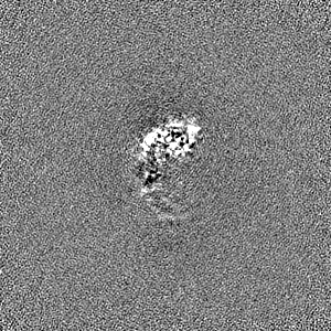
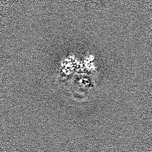
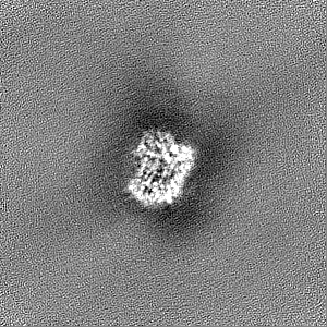
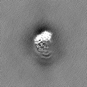
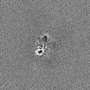
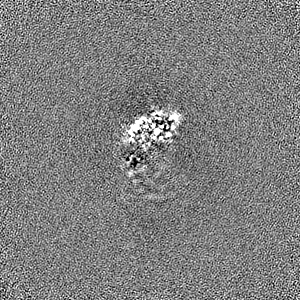
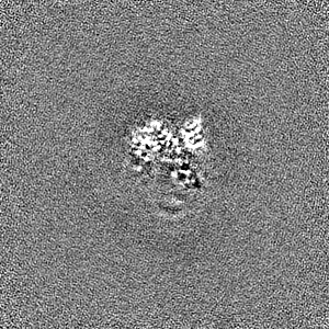
ジャーナル: Sci Rep / 年: 2023 タイトル: Cryo-EM structure of SKP1-SKP2-CKS1 in complex with CDK2-cyclin A-p27KIP1. 著者: Rhianna J Rowland / Richard Heath / Daniel Maskell / Rebecca F Thompson / Neil A Ranson / James N Blaza / Jane A Endicott / Martin E M Noble / Marco Salamina / 要旨: p27KIP1 (cyclin-dependent kinase inhibitor 1B, p27) is a member of the CIP/KIP family of CDK (cyclin dependent kinase) regulators that inhibit cell cycle CDKs. p27 phosphorylation by CDK1/2, signals ...p27KIP1 (cyclin-dependent kinase inhibitor 1B, p27) is a member of the CIP/KIP family of CDK (cyclin dependent kinase) regulators that inhibit cell cycle CDKs. p27 phosphorylation by CDK1/2, signals its recruitment to the SCF (S-phase kinase associated protein 1 (SKP1)-cullin-SKP2) E3 ubiquitin ligase complex for proteasomal degradation. The nature of p27 binding to SKP2 and CKS1 was revealed by the SKP1-SKP2-CKS1-p27 phosphopeptide crystal structure. Subsequently, a model for the hexameric CDK2-cyclin A-CKS1-p27-SKP1-SKP2 complex was proposed by overlaying an independently determined CDK2-cyclin A-p27 structure. Here we describe the experimentally determined structure of the isolated CDK2-cyclin A-CKS1-p27-SKP1-SKP2 complex at 3.4 Å global resolution using cryogenic electron microscopy. This structure supports previous analysis in which p27 was found to be structurally dynamic, transitioning from disordered to nascent secondary structure on target binding. We employed 3D variability analysis to further explore the conformational space of the hexameric complex and uncovered a previously unidentified hinge motion centred on CKS1. This flexibility gives rise to open and closed conformations of the hexameric complex that we propose may contribute to p27 regulation by facilitating recognition with SCF. This 3D variability analysis further informed particle subtraction and local refinement approaches to enhance the local resolution of the complex.
 ムービー
ムービー コントローラー


 データを開く
 基本情報
基本情報
 マップデータ
 試料
試料 キーワード
キーワード cell cycle (細胞周期) /
  機能・相同性情報
 データ登録者
 英国, 1件
  引用
引用 構造の表示
構造の表示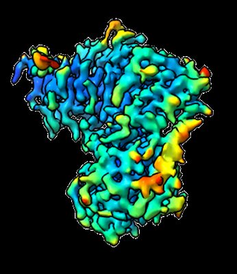
 ダウンロードとリンク
 emd_16344.png
 http://ftp.pdbj.org/pub/emdb/structures/EMD-16344


 Z
Z Y
Y X
X


 試料の構成要素
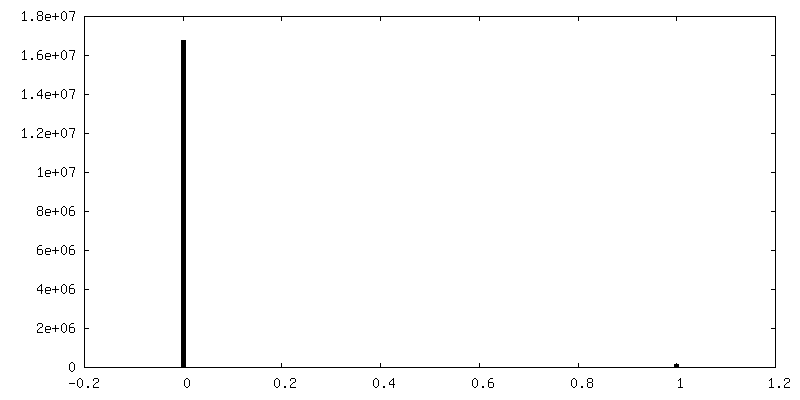
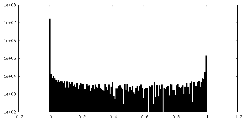
 解析
解析 電子顕微鏡法