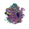
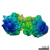
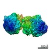
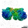
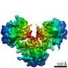
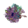
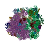
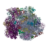
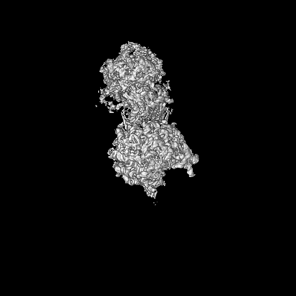
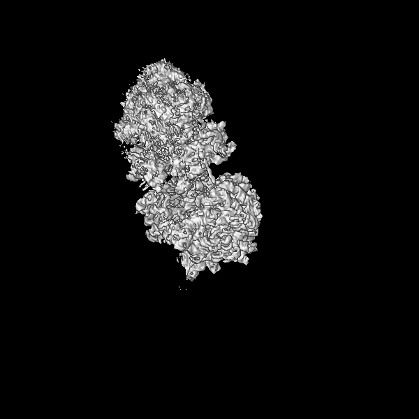
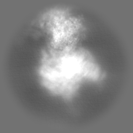
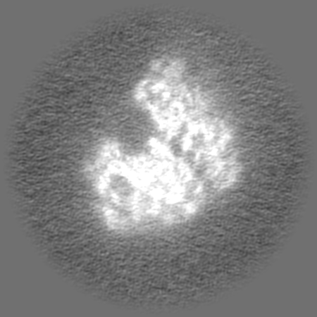
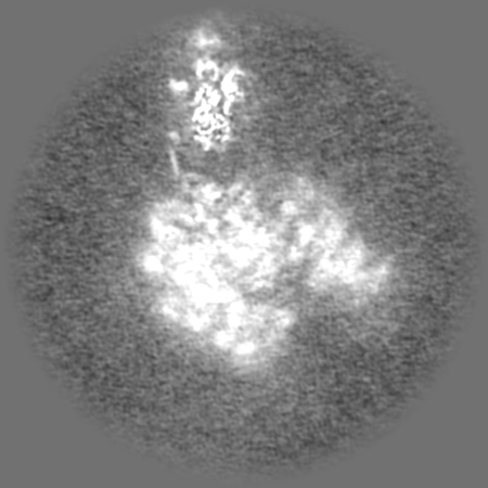
- EMDB-13958: Structure of the SmrB-bound E. coli disome - collided 70S ribosome -
+
データを開く
IDまたはキーワード:
読み込み中...
-
基本情報
登録情報
データベース: EMDB / ID: EMD-13958
タイトル
Structure of the SmrB-bound E. coli disome - collided 70S ribosome
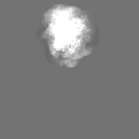
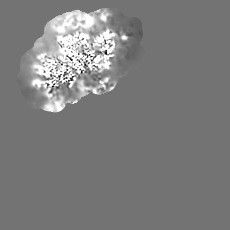
マップデータ
disome map
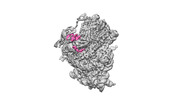
試料
複合体: collided ribosome of the SmrB-bound disome structure
RNA: x 6種
タンパク質・ペプチド: x 53種
リガンド: x 3種
機能・相同性
機能・相同性情報
stringent response / mRNA base-pairing translational repressor activity / ornithine decarboxylase inhibitor activity / transcription antitermination factor activity, RNA binding / transcriptional attenuation / endoribonuclease inhibitor activity / RNA-binding transcription regulator activity / negative regulation of cytoplasmic translation / translational termination / DnaA-L2 complex ...stringent response / mRNA base-pairing translational repressor activity / ornithine decarboxylase inhibitor activity / transcription antitermination factor activity, RNA binding / transcriptional attenuation / endoribonuclease inhibitor activity / RNA-binding transcription regulator activity / negative regulation of cytoplasmic translation / translational termination / DnaA-L2 complex / four-way junction DNA binding / translation repressor activity / negative regulation of DNA-templated DNA replication initiation / ribosome assembly / mRNA regulatory element binding translation repressor activity / assembly of large subunit precursor of preribosome / transcription elongation factor complex / response to reactive oxygen species / DNA endonuclease activity / cytosolic ribosome assembly / regulation of DNA-templated transcription elongation / transcription antitermination / DNA-templated transcription termination / response to radiation / mRNA 5'-UTR binding / ribosomal large subunit assembly / small ribosomal subunit rRNA binding / ribosomal small subunit assembly / cytosolic small ribosomal subunit / large ribosomal subunit rRNA binding / large ribosomal subunit / リボソーム生合成 / regulation of translation / small ribosomal subunit / cytoplasmic translation / 5S rRNA binding / cytosolic large ribosomal subunit / transferase activity / endonuclease activity / tRNA binding / negative regulation of translation / rRNA binding / リボソーム / structural constituent of ribosome / ribonucleoprotein complex / 翻訳 (生物学) / response to antibiotic / mRNA binding / negative regulation of DNA-templated transcription / DNA binding / RNA binding / zinc ion binding / 生体膜 / metal ion binding / 細胞質基質 / 細胞質 類似検索 - 分子機能
Uncharacterised protein family UPF0115 / Smr domain superfamily / Smr domain / Small MutS-related domain / Smr domain / Smr domain profile. / Ribosomal protein S1 / Ribosomal protein S1-like / RNA-binding domain, S1 / Ribosomal protein L1, bacterial-type ...Uncharacterised protein family UPF0115 / Smr domain superfamily / Smr domain / Small MutS-related domain / Smr domain / Smr domain profile. / Ribosomal protein S1 / Ribosomal protein S1-like / RNA-binding domain, S1 / Ribosomal protein L1, bacterial-type / S1 domain profile. / Ribosomal protein L1, conserved site / Ribosomal protein L1 / Ribosomal protein L1 signature. / Ribosomal protein L1, 3-layer alpha/beta-sandwich / Ribosomal protein S21, conserved site / Ribosomal protein S21 signature. / Ribosomal protein L25, short-form / Ribosomal protein L1-like / Ribosomal protein L1/ribosomal biogenesis protein / Ribosomal protein S14, bacterial/plastid / Ribosomal protein L1p/L10e family / Ribosomal protein S1-like RNA-binding domain / Ribosomal protein L11, bacterial-type / S1 RNA binding domain / Ribosomal protein L31 type A / Ribosomal protein S21 superfamily / S1 domain / Ribosomal protein S21 / Ribosomal protein S16, conserved site / Ribosomal protein S16 signature. / Ribosomal protein S21 / Ribosomal protein L31 signature. / Ribosomal protein L31 / Ribosomal protein L11, conserved site / Ribosomal protein L31 superfamily / Ribosomal protein L31 / Ribosomal protein L21, conserved site / Ribosomal protein L21 signature. / Ribosomal protein L11 signature. / Ribosomal protein L16 signature 1. / Ribosomal protein L6, conserved site / Ribosomal protein L6 signature 1. / Ribosomal protein L16, conserved site / Ribosomal protein L16 signature 2. / Ribosomal protein L11, N-terminal / Ribosomal protein L9 signature. / Ribosomal protein L17 signature. / Ribosomal protein L11/L12 / Ribosomal protein L9, bacteria/chloroplast / Ribosomal protein L11, C-terminal / Ribosomal protein L11, C-terminal domain superfamily / Ribosomal protein L11/L12, N-terminal domain superfamily / Ribosomal protein L11/L12 / Ribosomal protein L9, C-terminal / Ribosomal protein L9, C-terminal domain / Ribosomal protein L9, C-terminal domain superfamily / Ribosomal protein L11, N-terminal domain / Ribosomal protein L11, RNA binding domain / Ribosomal L25p family / Ribosomal protein L25 / Ribosomal protein L28/L24 superfamily / Ribosomal protein L25/Gln-tRNA synthetase, N-terminal / Ribosomal protein L32p, bacterial type / Ribosomal protein L25/Gln-tRNA synthetase, anti-codon-binding domain superfamily / Ribosomal protein L9, N-terminal domain superfamily / Ribosomal protein L9 / Ribosomal protein L9, N-terminal / Ribosomal protein L9, N-terminal domain / Ribosomal protein L28 / Ribosomal protein L35, conserved site / Ribosomal protein L35 signature. / Ribosomal protein L33, conserved site / Ribosomal protein L33 signature. / Ribosomal protein L35, non-mitochondrial / Ribosomal protein L5, bacterial-type / Ribosomal protein L6, bacterial-type / Ribosomal protein L18, bacterial-type / Ribosomal protein L19, conserved site / Ribosomal protein L19 signature. / Ribosomal protein L9/RNase H1, N-terminal / Ribosomal protein S3, bacterial-type / Ribosomal protein S6, conserved site / Ribosomal protein S6 signature. / Ribosomal protein L20 signature. / Ribosomal protein S19, bacterial-type / Ribosomal protein L27, conserved site / Ribosomal protein L27 signature. / Ribosomal protein S7, bacterial/organellar-type / Ribosomal protein S11, bacterial-type / Ribosomal protein S13, bacterial-type / Ribosomal protein S20 / Ribosomal protein S20 superfamily / Ribosomal protein S20 / Ribosomal protein S9, bacterial/plastid / Ribosomal protein S4, bacterial-type / 30S ribosomal protein S17 / Ribosomal protein S5, bacterial-type / Ribosomal protein L14P, bacterial-type / Ribosomal protein L34, conserved site 類似検索 - ドメイン・相同性
Small ribosomal subunit protein uS14 / 50S ribosomal protein L31 / 30S ribosomal protein S9 / 30S ribosomal protein S18 / 30S ribosomal protein S5 / 50S ribosomal protein L1 / Small ribosomal subunit protein uS13 / 30S ribosomal protein S17 / 30S ribosomal protein S11 / Large ribosomal subunit protein uL23 ...Small ribosomal subunit protein uS14 / 50S ribosomal protein L31 / 30S ribosomal protein S9 / 30S ribosomal protein S18 / 30S ribosomal protein S5 / 50S ribosomal protein L1 / Small ribosomal subunit protein uS13 / 30S ribosomal protein S17 / 30S ribosomal protein S11 / Large ribosomal subunit protein uL23 / Large ribosomal subunit protein bL17 / 30S ribosomal protein S1 / 50S ribosomal protein L3 / Small ribosomal subunit protein uS12 / Small ribosomal subunit protein uS8 / Small ribosomal subunit protein uS4 / Small ribosomal subunit protein bS6 / Small ribosomal subunit protein uS7 / Large ribosomal subunit protein uL15 / Large ribosomal subunit protein uL11 / Large ribosomal subunit protein bL19 / Large ribosomal subunit protein bL20 / Large ribosomal subunit protein bL28 / Large ribosomal subunit protein uL29 / Large ribosomal subunit protein bL32 / Large ribosomal subunit protein bL33 / Large ribosomal subunit protein bL34 / Large ribosomal subunit protein bL35 / Large ribosomal subunit protein bL9 / Small ribosomal subunit protein uS10 / Small ribosomal subunit protein bS16 / Small ribosomal subunit protein bS20 / Small ribosomal subunit protein uS2 / Small ribosomal subunit protein uS3 / Large ribosomal subunit protein uL13 / Large ribosomal subunit protein uL14 / Large ribosomal subunit protein uL16 / Small ribosomal subunit protein uS15 / Large ribosomal subunit protein bL21 / Large ribosomal subunit protein uL30 / Large ribosomal subunit protein uL6 / Large ribosomal subunit protein uL18 / Large ribosomal subunit protein uL2 / Large ribosomal subunit protein uL24 / Large ribosomal subunit protein uL4 / Large ribosomal subunit protein uL22 / Large ribosomal subunit protein uL5 / Small ribosomal subunit protein bS21 / Large ribosomal subunit protein bL25 / UPF0115 protein YfcN / Small ribosomal subunit protein uS19 / Large ribosomal subunit protein bL27 類似検索 - 構成要素
ジャーナル: Nature / 年: 2022 タイトル: Ribosome collisions induce mRNA cleavage and ribosome rescue in bacteria. 著者: Kazuki Saito / Hanna Kratzat / Annabelle Campbell / Robert Buschauer / A Maxwell Burroughs / Otto Berninghausen / L Aravind / Rachel Green / Roland Beckmann / Allen R Buskirk / 要旨: Ribosome rescue pathways recycle stalled ribosomes and target problematic mRNAs and aborted proteins for degradation. In bacteria, it remains unclear how rescue pathways distinguish ribosomes stalled ...Ribosome rescue pathways recycle stalled ribosomes and target problematic mRNAs and aborted proteins for degradation. In bacteria, it remains unclear how rescue pathways distinguish ribosomes stalled in the middle of a transcript from actively translating ribosomes. Here, using a genetic screen in Escherichia coli, we discovered a new rescue factor that has endonuclease activity. SmrB cleaves mRNAs upstream of stalled ribosomes, allowing the ribosome rescue factor tmRNA (which acts on truncated mRNAs) to rescue upstream ribosomes. SmrB is recruited to ribosomes and is activated by collisions. Cryo-electron microscopy structures of collided disomes from E. coli and Bacillus subtilis show distinct and conserved arrangements of individual ribosomes and the composite SmrB-binding site. These findings reveal the underlying mechanisms by which ribosome collisions trigger ribosome rescue in bacteria.
 ムービー
ムービー コントローラー


 データを開く
 基本情報
基本情報
 マップデータ
 試料
試料 機能・相同性情報
 stringent response / mRNA base-pairing translational repressor activity / ornithine decarboxylase inhibitor activity / transcription antitermination factor activity, RNA binding /
 データ登録者
 ドイツ, 1件
  引用
引用
 構造の表示
構造の表示
 ダウンロードとリンク
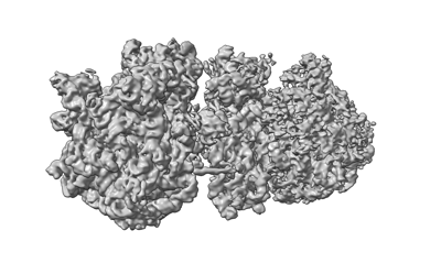 emd_13958.png
 http://ftp.pdbj.org/pub/emdb/structures/EMD-13958
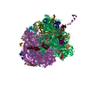


 Z (Sec.)
 Y (Row.)
 X (Col.)


 試料の構成要素
 解析
解析 電子顕微鏡法