 ムービー
ムービー コントローラー


+ データを開く

- 基本情報
基本情報
| 登録情報 | データベース: EMDB / ID: EMD-6489 | |||||||||
|---|---|---|---|---|---|---|---|---|---|---|
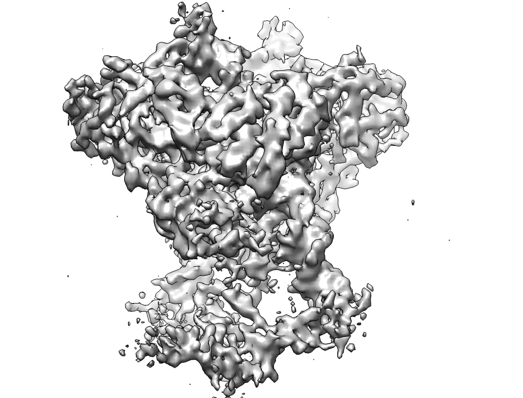
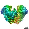
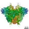
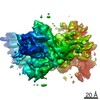
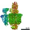
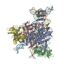
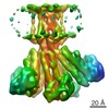
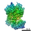
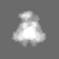
| タイトル | Cryo-electron microscopy structure of RAG PC (with NBD, no symmetry) | |||||||||
 マップデータ マップデータ | Reconstruction of RAG PC without symmetry, showing NBD | |||||||||
 試料 試料 |
| |||||||||
 キーワード キーワード |  RAG1 / RAG2 / V(D)J recombination (V(D)J遺伝子再構成) / Paired complex / Antigen receptor recombination / T and B cell development RAG1 / RAG2 / V(D)J recombination (V(D)J遺伝子再構成) / Paired complex / Antigen receptor recombination / T and B cell development | |||||||||
| 機能・相同性 |  機能・相同性情報 機能・相同性情報somatic diversification of immune receptors via germline recombination within a single locus / hematopoietic or lymphoid organ development / protein binding / protein-DNA complex assembly / DNA recombinase complex / endodeoxyribonuclease complex / lymphocyte differentiation / immunoglobulin V(D)J recombination / V(D)J遺伝子再構成 / phosphatidylinositol-3,4-bisphosphate binding ...somatic diversification of immune receptors via germline recombination within a single locus / hematopoietic or lymphoid organ development / protein binding / protein-DNA complex assembly / DNA recombinase complex / endodeoxyribonuclease complex / lymphocyte differentiation / immunoglobulin V(D)J recombination / V(D)J遺伝子再構成 / phosphatidylinositol-3,4-bisphosphate binding / phosphatidylinositol-3,5-bisphosphate binding / phosphatidylinositol-3,4,5-trisphosphate binding / T cell differentiation / methylated histone binding / phosphatidylinositol-4,5-bisphosphate binding / phosphatidylinositol binding / B cell differentiation / thymus development / RING-type E3 ubiquitin transferase / ubiquitin-protein transferase activity / ubiquitin protein ligase activity / T cell differentiation in thymus / chromatin organization / histone binding / endonuclease activity / DNA recombination / sequence-specific DNA binding / 獲得免疫系 / 加水分解酵素; エステル加水分解酵素 / chromatin binding / magnesium ion binding / protein homodimerization activity / DNA binding / zinc ion binding / metal ion binding / 細胞核類似検索 - 分子機能 | |||||||||
| 生物種 |  Danio rerio (ゼブラフィッシュ) Danio rerio (ゼブラフィッシュ) | |||||||||
| 手法 | 単粒子再構成法 / クライオ電子顕微鏡法 / 解像度: 4.6 Å | |||||||||
 データ登録者 データ登録者 | Ru H / Chambers MG / Fu T / Tong AB / Liao M / Wu H | |||||||||
 引用 引用 | ジャーナル: Cell / 年: 2015 タイトル: Molecular Mechanism of V(D)J Recombination from Synaptic RAG1-RAG2 Complex Structures. 著者: Heng Ru / Melissa G Chambers / Tian-Min Fu / Alexander B Tong / Maofu Liao / Hao Wu /  要旨: Diverse repertoires of antigen-receptor genes that result from combinatorial splicing of coding segments by V(D)J recombination are hallmarks of vertebrate immunity. The (RAG1-RAG2)2 recombinase (RAG) ...Diverse repertoires of antigen-receptor genes that result from combinatorial splicing of coding segments by V(D)J recombination are hallmarks of vertebrate immunity. The (RAG1-RAG2)2 recombinase (RAG) recognizes recombination signal sequences (RSSs) containing a heptamer, a spacer of 12 or 23 base pairs, and a nonamer (12-RSS or 23-RSS) and introduces precise breaks at RSS-coding segment junctions. RAG forms synaptic complexes only with one 12-RSS and one 23-RSS, a dogma known as the 12/23 rule that governs the recombination fidelity. We report cryo-electron microscopy structures of synaptic RAG complexes at up to 3.4 Å resolution, which reveal a closed conformation with base flipping and base-specific recognition of RSSs. Distortion at RSS-coding segment junctions and base flipping in coding segments uncover the two-metal-ion catalytic mechanism. Induced asymmetry involving tilting of the nonamer-binding domain dimer of RAG1 upon binding of HMGB1-bent 12-RSS or 23-RSS underlies the molecular mechanism for the 12/23 rule. | |||||||||
| 履歴 |
|
- 構造の表示
構造の表示
| ムービー |
ムービービューア |
|---|---|
| 構造ビューア | EMマップ: SurfViewMolmilJmol/JSmol |
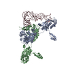
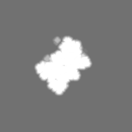
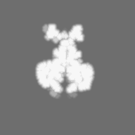
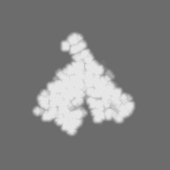
| 添付画像 |
- ダウンロードとリンク
-EMDBアーカイブ
| マップデータ | emd_6489.map.gz | 25.3 MB | EMDBマップデータ形式 | |
|---|---|---|---|---|
| ヘッダ (付随情報) | emd-6489-v30.xmlemd-6489.xml | 15.1 KB 15.1 KB | 表示 表示 | EMDBヘッダ |
| FSC (解像度算出) | emd_6489_fsc.xml | 6.7 KB | 表示 | FSCデータファイル |
| 画像 |  emd_6489.png emd_6489.png | 421.2 KB | ||
| マスクデータ | emd_6489_msk_1.map | 27 MB | マスクマップ | |
| アーカイブディレクトリ |  http://ftp.pdbj.org/pub/emdb/structures/EMD-6489ftp://ftp.pdbj.org/pub/emdb/structures/EMD-6489 http://ftp.pdbj.org/pub/emdb/structures/EMD-6489ftp://ftp.pdbj.org/pub/emdb/structures/EMD-6489 | HTTPS FTP |
-関連構造データ


-リンク
| EMDBのページ | EMDB (EBI/PDBe) / EMDataResource |
|---|---|
| 「今月の分子」の関連する項目 |


-マップ
| ファイル | ダウンロード / ファイル: emd_6489.map.gz / 形式: CCP4 / 大きさ: 26.4 MB / タイプ: IMAGE STORED AS FLOATING POINT NUMBER (4 BYTES) | ||||||||||||||||||||||||||||||||||||||||||||||||||||||||||||||||||||
|---|---|---|---|---|---|---|---|---|---|---|---|---|---|---|---|---|---|---|---|---|---|---|---|---|---|---|---|---|---|---|---|---|---|---|---|---|---|---|---|---|---|---|---|---|---|---|---|---|---|---|---|---|---|---|---|---|---|---|---|---|---|---|---|---|---|---|---|---|---|
| 注釈 | Reconstruction of RAG PC without symmetry, showing NBD | ||||||||||||||||||||||||||||||||||||||||||||||||||||||||||||||||||||
| ボクセルのサイズ | X=Y=Z: 1.23 Å | ||||||||||||||||||||||||||||||||||||||||||||||||||||||||||||||||||||
| 密度 |
| ||||||||||||||||||||||||||||||||||||||||||||||||||||||||||||||||||||
| 対称性 | 空間群: 1 | ||||||||||||||||||||||||||||||||||||||||||||||||||||||||||||||||||||
| 詳細 | EMDB XML:
CCP4マップ ヘッダ情報:
| ||||||||||||||||||||||||||||||||||||||||||||||||||||||||||||||||||||
-添付データ
-セグメンテーションマップ: This mask represents the full molecule
| 注釈 | This mask represents the full molecule | ||||||||||||
|---|---|---|---|---|---|---|---|---|---|---|---|---|---|
| ファイル | emd_6489_msk_1.map | ||||||||||||
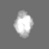
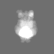
| 投影像・断面図 |
| ||||||||||||
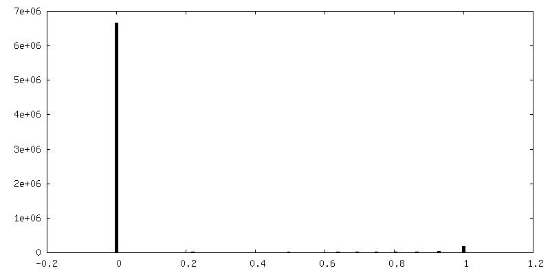
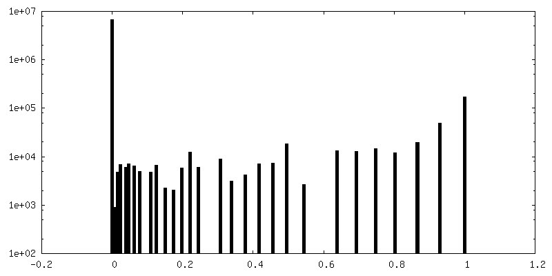
| 密度ヒストグラム |
 Z
Z Y
Y X
X

- 試料の構成要素
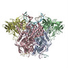
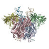
-全体 : Paired Complex of RAG1-RAG2
| 全体 | 名称: Paired Complex of RAG1-RAG2 |
|---|---|
| 要素 |
|
-超分子 #1000: Paired Complex of RAG1-RAG2
| 超分子 | 名称: Paired Complex of RAG1-RAG2 / タイプ: sample / ID: 1000 / 詳細: The sample was monodisperse. 集合状態: Dimer of RAG1-RAG2 binds to nicked 12-RSS and 23-RSS intermediates with HMGB1 Number unique components: 1 |
|---|---|
| 分子量 | 実験値: 400 KDa / 理論値: 400 KDa / 手法: Size exclusion chromatography |
-分子 #1: Recombination Activating Gene 1 and 2
| 分子 | 名称: Recombination Activating Gene 1 and 2 / タイプ: protein_or_peptide / ID: 1 / Name.synonym: RAG1 and RAG2 / コピー数: 2 / 集合状態: Dimer of RAG1-RAG2 / 組換発現: Yes |
|---|---|
| 由来(天然) | 生物種: Danio rerio (ゼブラフィッシュ) / 株: AB / 別称: Zebrafish / 細胞: B and T lymphocytes / Organelle: Nucleus |
| 分子量 | 実験値: 400 KDa / 理論値: 400 KDa |
| 組換発現 | 生物種:   Spodoptera frugiperda (ツマジロクサヨトウ) Spodoptera frugiperda (ツマジロクサヨトウ)組換細胞: Sf9 / 組換プラスミド: pFastBac1 |
| 配列 | UniProtKB: V(D)J recombination-activating protein 1 GO: DNA binding, endonuclease activity, ubiquitin-protein transferase activity, protein binding, zinc ion binding, DNA binding, chromatin binding, protein binding, phosphatidylinositol-4,5- ...GO: DNA binding, endonuclease activity, ubiquitin-protein transferase activity, protein binding, zinc ion binding, DNA binding, chromatin binding, protein binding, phosphatidylinositol-4,5-bisphosphate binding, phosphatidylinositol-3,4,5-trisphosphate bindingInterPro: V(D)J recombination-activating protein 1, RAG nonamer-binding domain, Zinc finger, C3HC4 RING-type, Zinc finger, RING-type, Zinc finger, RING/FYVE/PHD-type, Zinc finger, RING-type, ...InterPro: V(D)J recombination-activating protein 1, RAG nonamer-binding domain, Zinc finger, C3HC4 RING-type, Zinc finger, RING-type, Zinc finger, RING/FYVE/PHD-type, Zinc finger, RING-type, conserved site, Galactose oxidase/kelch, beta-propeller, Kelch-type beta propeller, V-D-J recombination activating protein 2, Recombination activating protein 2, PHD domain, Zinc finger, FYVE/PHD-type |
-実験情報
-構造解析
| 手法 | クライオ電子顕微鏡法 |
|---|---|
 解析 解析 | 単粒子再構成法 |
| 試料の集合状態 | particle |
-試料調製
| 濃度 | 0.4 mg/mL |
|---|---|
| 緩衝液 | pH: 7.5 / 詳細: 150 mM NaCl, 20 mM HEPES, 10 mM CaCl2, 1 mM TCEP |
| グリッド | 詳細: 400 mesh Quantifoil holey carbon grid, glow discharged |
| 凍結 | 凍結剤: ETHANE / チャンバー内湿度: 85 % / チャンバー内温度: 120 K / 装置: GATAN CRYOPLUNGE 3 / 手法: Blot for 2.5 seconds before plunging. |
- 電子顕微鏡法
| 顕微鏡 | FEI POLARA 300 |
|---|---|
| 電子線 | 加速電圧: 300 kV / 電子線源: FIELD EMISSION GUN |
| 電子光学系 | 倍率(補正後): 40607 / 照射モード: FLOOD BEAM / 撮影モード: BRIGHT FIELDBright-field microscopy / Cs: 2.0 mm / 最大 デフォーカス(公称値): 2.5 µm / 最小 デフォーカス(公称値): 1.5 µm / 倍率(公称値): 31000 |
| 試料ステージ | 試料ホルダーモデル: GATAN LIQUID NITROGEN |
| 温度 | 最低: 80 K / 最高: 105 K / 平均: 100 K |
| アライメント法 | Legacy - 非点収差: Objective lens astigmatism was corrected at 150,000 times magnification |
| 日付 | 2015年3月10日 |
| 撮影 | カテゴリ: CCD / フィルム・検出器のモデル: GATAN K2 (4k x 4k) / 実像数: 550 / 平均電子線量: 41 e/Å2 詳細: Every image is the average of 30 frames recorded by the direct electron detector. ビット/ピクセル: 8 |
| 実験機器 |  モデル: Tecnai Polara / 画像提供: FEI Company |
-画像解析
| CTF補正 | 詳細: Each particle |
|---|---|
| 最終 再構成 | アルゴリズム: OTHER / 解像度のタイプ: BY AUTHOR / 解像度: 4.6 Å / 解像度の算出法: OTHER / ソフトウェア - 名称: SPIDER, Relion / 使用した粒子像数: 14129 |
| 詳細 | Image processing was carried out using SAMUEL and Relion. |
| FSC曲線 (解像度の算出) |  |
