 Movie
Movie Controller

[English] 日本語
 Yorodumi
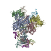
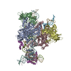
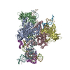
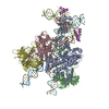
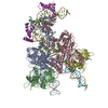
- PDB-3jbw: Cryo-electron microscopy structure of RAG Paired Complex (with NB... -
+ Open data

- Basic information
| Entry | Database: PDB / ID: 3jbw | ||||||
|---|---|---|---|---|---|---|---|
| Title | Cryo-electron microscopy structure of RAG Paired Complex (with NBD, no symmetry) | ||||||
 Components Components |
| ||||||
 Keywords Keywords | RECOMBINATION/DNA / RAG1 / RAG2 / V(D)J recombination / Paired complex / Antigen receptor gene recombination / T and B cell development / RECOMBINATION-DNA complex | ||||||
| Function / homology |  Function and homology information Function and homology informationsomatic diversification of immune receptors via germline recombination within a single locus / hematopoietic or lymphoid organ development / protein-DNA complex assembly / DNA recombinase complex / endodeoxyribonuclease complex / lymphocyte differentiation / pre-B cell allelic exclusion / immunoglobulin V(D)J recombination / V(D)J recombination / phosphatidylinositol-3,4-bisphosphate binding ...somatic diversification of immune receptors via germline recombination within a single locus / hematopoietic or lymphoid organ development / protein-DNA complex assembly / DNA recombinase complex / endodeoxyribonuclease complex / lymphocyte differentiation / pre-B cell allelic exclusion / immunoglobulin V(D)J recombination / V(D)J recombination / phosphatidylinositol-3,4-bisphosphate binding / histone H3K4me3 reader activity / phosphatidylinositol-3,5-bisphosphate binding / phosphatidylinositol-3,4,5-trisphosphate binding / T cell differentiation / phosphatidylinositol-4,5-bisphosphate binding / B cell differentiation / thymus development / phosphatidylinositol binding / RING-type E3 ubiquitin transferase / ubiquitin-protein transferase activity / T cell differentiation in thymus / ubiquitin protein ligase activity / chromatin organization / endonuclease activity / histone binding / sequence-specific DNA binding / Hydrolases; Acting on ester bonds / adaptive immune response / hydrolase activity / chromatin binding / magnesium ion binding / protein homodimerization activity / zinc ion binding / metal ion binding / nucleus Similarity search - Function | ||||||
| Biological species |   Homo sapiens (human) Homo sapiens (human) | ||||||
| Method | ELECTRON MICROSCOPY / single particle reconstruction / cryo EM / Resolution: 4.6 Å | ||||||
 Authors Authors | Ru, H. / Chambers, M.G. / Fu, T.-M. / Tong, A.B. / Liao, M. / Wu, H. | ||||||
 Citation Citation | Journal: Cell / Year: 2015 Title: Molecular Mechanism of V(D)J Recombination from Synaptic RAG1-RAG2 Complex Structures. Authors: Heng Ru / Melissa G Chambers / Tian-Min Fu / Alexander B Tong / Maofu Liao / Hao Wu /  Abstract: Diverse repertoires of antigen-receptor genes that result from combinatorial splicing of coding segments by V(D)J recombination are hallmarks of vertebrate immunity. The (RAG1-RAG2)2 recombinase (RAG) ...Diverse repertoires of antigen-receptor genes that result from combinatorial splicing of coding segments by V(D)J recombination are hallmarks of vertebrate immunity. The (RAG1-RAG2)2 recombinase (RAG) recognizes recombination signal sequences (RSSs) containing a heptamer, a spacer of 12 or 23 base pairs, and a nonamer (12-RSS or 23-RSS) and introduces precise breaks at RSS-coding segment junctions. RAG forms synaptic complexes only with one 12-RSS and one 23-RSS, a dogma known as the 12/23 rule that governs the recombination fidelity. We report cryo-electron microscopy structures of synaptic RAG complexes at up to 3.4 Å resolution, which reveal a closed conformation with base flipping and base-specific recognition of RSSs. Distortion at RSS-coding segment junctions and base flipping in coding segments uncover the two-metal-ion catalytic mechanism. Induced asymmetry involving tilting of the nonamer-binding domain dimer of RAG1 upon binding of HMGB1-bent 12-RSS or 23-RSS underlies the molecular mechanism for the 12/23 rule. | ||||||
| History |
|
- Structure visualization
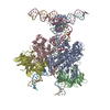
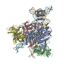
| Movie |
Movie viewer |
|---|---|
| Structure viewer | Molecule: MolmilJmol/JSmol |
- Downloads & links
-Download
| PDBx/mmCIF format | 3jbw.cif.gz | 1 MB | Display | PDBx/mmCIF format |
|---|---|---|---|---|
| PDB format | pdb3jbw.ent.gz | 872.5 KB | Display | PDB format |
| PDBx/mmJSON format | 3jbw.json.gz | Tree view | PDBx/mmJSON format | |
| Others |  Other downloads Other downloads |
-Validation report
| Arichive directory | https://data.pdbj.org/pub/pdb/validation_reports/jb/3jbwftp://data.pdbj.org/pub/pdb/validation_reports/jb/3jbw | HTTPS FTP |
|---|
-Related structure data
| Related structure data | 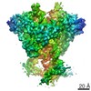 6489MC 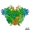 6487C 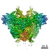 6488C 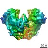 6490C 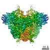 6491C 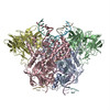 3jbxC 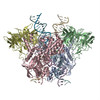 3jbyC M: map data used to model this data C: citing same article ( |
|---|---|
| Similar structure data |

-Links
 PDBj
PDBj


- Assembly
| Deposited unit | 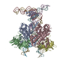
|
|---|---|
| 1 |
|
-Components
-V(D)J recombination-activating protein ... , 2 types, 4 molecules ACBD
| #1: Protein | Mass: 87684.125 Da / Num. of mol.: 2 Source method: isolated from a genetically manipulated source Source: (gene. exp.)   Spodoptera frugiperda (fall armyworm) Spodoptera frugiperda (fall armyworm)References: UniProt: O13033, Hydrolases; Acting on ester bonds, Ligases; Forming carbon-nitrogen bonds; Acid-amino-acid ligases (peptide synthases) #2: Protein | Mass: 59435.930 Da / Num. of mol.: 2 Source method: isolated from a genetically manipulated source Source: (gene. exp.) Spodoptera frugiperda (fall armyworm) / References: UniProt: Q1RLW7, UniProt: O13034*PLUS |
|---|
-DNA chain , 3 types, 4 molecules EFIJ
| #3: DNA chain | Mass: 10439.762 Da / Num. of mol.: 1 / Source method: obtained synthetically / Source: (synth.) Homo sapiens (human) |
|---|---|
| #4: DNA chain | Mass: 15439.880 Da / Num. of mol.: 1 / Source method: obtained synthetically / Source: (synth.) Homo sapiens (human) |
| #7: DNA chain | Mass: 4880.164 Da / Num. of mol.: 2 / Source method: obtained synthetically / Details: Coding end DNA forward strand / Source: (synth.) Homo sapiens (human) |
-Nicked 23-RSS intermediate ... , 2 types, 2 molecules GH
| #5: DNA chain | Mass: 18870.094 Da / Num. of mol.: 1 / Source method: obtained synthetically / Source: (synth.) Homo sapiens (human) |
|---|---|
| #6: DNA chain | Mass: 13806.871 Da / Num. of mol.: 1 / Source method: obtained synthetically / Source: (synth.) Homo sapiens (human) |
-Non-polymers , 1 types, 2 molecules 
| #8: Chemical |  Mass: 65.409 Da / Num. of mol.: 2 / Source method: obtained synthetically / Formula: Zn Mass: 65.409 Da / Num. of mol.: 2 / Source method: obtained synthetically / Formula: Zn |
|---|
-Experimental details
-Experiment
| Experiment | Method: ELECTRON MICROSCOPY |
|---|---|
| EM experiment | Aggregation state: PARTICLE / 3D reconstruction method: single particle reconstruction |
- Sample preparation
| Component |
| ||||||||||||||||||||
|---|---|---|---|---|---|---|---|---|---|---|---|---|---|---|---|---|---|---|---|---|---|
| Molecular weight | Value: 0.4 MDa / Experimental value: YES | ||||||||||||||||||||
| Buffer solution | Name: Polymix buffer / pH: 7.5 / Details: 150 mM NaCl, 20 mM HEPES, 10 mM CaCl2, 1 mM TCEP | ||||||||||||||||||||
| Specimen | Conc.: 0.4 mg/ml / Embedding applied: NO / Shadowing applied: NO / Staining applied: NO / Vitrification applied: YES | ||||||||||||||||||||
| Specimen support | Details: 400 mesh Quantifoil holey carbon grid, glow discharged | ||||||||||||||||||||
| Vitrification | Instrument: GATAN CRYOPLUNGE 3 / Cryogen name: ETHANE / Temp: 120 K / Humidity: 85 % Details: Blot for 2.5 seconds before plunging into liquid ethane (GATAN CRYOPLUNGE 3). Method: Blot for 2.5 seconds before plunging. |
- Electron microscopy imaging
| Experimental equipment |  Model: Tecnai Polara / Image courtesy: FEI Company |
|---|---|
| Microscopy | Model: FEI POLARA 300 / Date: Mar 10, 2015 |
| Electron gun | Electron source:  FIELD EMISSION GUN / Accelerating voltage: 300 kV / Illumination mode: FLOOD BEAM FIELD EMISSION GUN / Accelerating voltage: 300 kV / Illumination mode: FLOOD BEAM |
| Electron lens | Mode: BRIGHT FIELD / Nominal magnification: 31000 X / Calibrated magnification: 40607 X / Nominal defocus max: 2500 nm / Nominal defocus min: 1500 nm / Cs: 2 mm Astigmatism: Objective lens astigmatism was corrected at 150,000 times magnification. |
| Specimen holder | Specimen holder model: GATAN LIQUID NITROGEN / Temperature: 100 K / Temperature (max): 105 K / Temperature (min): 80 K |
| Image recording | Electron dose: 41 e/Å2 / Film or detector model: GATAN K2 (4k x 4k) |
| Image scans | Num. digital images: 550 |
- Processing
| EM software |
| ||||||||||||||||||||||||||||||||||||||||||||||||||||||||||||||||||||||||||||||
|---|---|---|---|---|---|---|---|---|---|---|---|---|---|---|---|---|---|---|---|---|---|---|---|---|---|---|---|---|---|---|---|---|---|---|---|---|---|---|---|---|---|---|---|---|---|---|---|---|---|---|---|---|---|---|---|---|---|---|---|---|---|---|---|---|---|---|---|---|---|---|---|---|---|---|---|---|---|---|---|
| CTF correction | Details: Each particle | ||||||||||||||||||||||||||||||||||||||||||||||||||||||||||||||||||||||||||||||
| Symmetry | Point symmetry: C1 (asymmetric) | ||||||||||||||||||||||||||||||||||||||||||||||||||||||||||||||||||||||||||||||
| 3D reconstruction | Method: Projection matching / Resolution: 4.6 Å / Resolution method: FSC 0.143 CUT-OFF / Num. of particles: 14129 / Nominal pixel size: 1.23 Å / Actual pixel size: 1.23 Å Details: (Single particle details: Image processing was carried out using SAMUEL and Relion.) (Single particle--Applied symmetry: C1) Symmetry type: POINT | ||||||||||||||||||||||||||||||||||||||||||||||||||||||||||||||||||||||||||||||
| Refinement | Resolution: 4.6→4.6 Å / SU ML: 1.64 / σ(F): 0 / Phase error: 62.82 / Stereochemistry target values: MLHL
| ||||||||||||||||||||||||||||||||||||||||||||||||||||||||||||||||||||||||||||||
| Solvent computation | Shrinkage radii: 0.9 Å / VDW probe radii: 1.11 Å / Solvent model: FLAT BULK SOLVENT MODEL | ||||||||||||||||||||||||||||||||||||||||||||||||||||||||||||||||||||||||||||||
| Displacement parameters | Biso max: 587.07 Å2 / Biso mean: 180.3618 Å2 / Biso min: 19.35 Å2 | ||||||||||||||||||||||||||||||||||||||||||||||||||||||||||||||||||||||||||||||
| Refinement step | Cycle: LAST / Resolution: 4.631→236.16 Å
| ||||||||||||||||||||||||||||||||||||||||||||||||||||||||||||||||||||||||||||||
| Refine LS restraints |
| ||||||||||||||||||||||||||||||||||||||||||||||||||||||||||||||||||||||||||||||
| LS refinement shell | Refine-ID: ELECTRON MICROSCOPY / Total num. of bins used: 4 / % reflection obs: 100 %
| ||||||||||||||||||||||||||||||||||||||||||||||||||||||||||||||||||||||||||||||
| Refinement TLS params. | Method: refined / Origin x: 113.7436 Å / Origin y: 115.0465 Å / Origin z: 119.5786 Å
| ||||||||||||||||||||||||||||||||||||||||||||||||||||||||||||||||||||||||||||||
| Refinement TLS group |
|
