Movie
Movie Controller

[English] 日本語
 Yorodumi
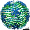
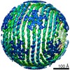
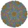
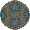
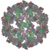
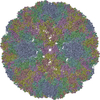
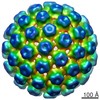
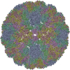
- PDB-6hy0: Atomic models of P1, P4 C-terminal fragment and P8 fitted in the ... -
+ Open data

- Basic information
| Entry | Database: PDB / ID: 6hy0 | ||||||
|---|---|---|---|---|---|---|---|
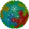
| Title | Atomic models of P1, P4 C-terminal fragment and P8 fitted in the bacteriophage phi6 nucleocapsid reconstructed with icosahedral symmetry | ||||||
 Components Components |
| ||||||
 Keywords Keywords |  VIRUS / dsRNA / capsid VIRUS / dsRNA / capsid | ||||||
| Function / homology | : / Major inner capsid protein P1 / ATPase P4 of dsRNA bacteriophage phi-12 / T=2 icosahedral viral capsid / viral inner capsid / viral nucleocapsid / RNA binding / identical protein binding / Major inner protein P1 Function and homology information Function and homology information | ||||||
| Biological species |  Pseudomonas phage phi6 (bacteriophage) Pseudomonas phage phi6 (bacteriophage) | ||||||
| Method | ELECTRON MICROSCOPY / single particle reconstruction / cryo EM / Resolution: 3.5 Å | ||||||
 Authors Authors | El Omari, K. / Ilca, S.L. / Stuart, D.I. / Huiskonen, J.T. | ||||||
 Citation Citation | Journal: Nature / Year: 2019 Title: Multiple liquid crystalline geometries of highly compacted nucleic acid in a dsRNA virus. Authors: Serban L Ilca / Xiaoyu Sun / Kamel El Omari / Abhay Kotecha / Felix de Haas / Frank DiMaio / Jonathan M Grimes / David I Stuart / Minna M Poranen / Juha T Huiskonen /     Abstract: Characterizing the genome of mature virions is pivotal to understanding the highly dynamic processes of virus assembly and infection. Owing to the different cellular fates of DNA and RNA, the life ...Characterizing the genome of mature virions is pivotal to understanding the highly dynamic processes of virus assembly and infection. Owing to the different cellular fates of DNA and RNA, the life cycles of double-stranded (ds)DNA and dsRNA viruses are dissimilar. In terms of nucleic acid packing, dsDNA viruses, which lack genome segmentation and intra-capsid transcriptional machinery, predominantly display single-spooled genome organizations. Because the release of dsRNA into the cytoplasm triggers host defence mechanisms, dsRNA viruses retain their genomes within a core particle that contains the enzymes required for RNA replication and transcription. The genomes of dsRNA viruses vary greatly in the degree of segmentation. In members of the Reoviridae family, genomes consist of 10-12 segments and exhibit a non-spooled arrangement mediated by RNA-dependent RNA polymerases. However, whether this arrangement is a general feature of dsRNA viruses remains unknown. Here, using cryo-electron microscopy to resolve the dsRNA genome structure of the tri-segmented bacteriophage ɸ6 of the Cystoviridae family, we show that dsRNA viruses can adopt a dsDNA-like single-spooled genome organization. We find that in this group of viruses, RNA-dependent RNA polymerases do not direct genome ordering, and the dsRNA can adopt multiple conformations. We build a model that encompasses 90% of the genome, and use this to quantify variation in the packing density and to characterize the different liquid crystalline geometries that are exhibited by the tightly compacted nucleic acid. Our results demonstrate that the canonical model for the packing of dsDNA can be extended to dsRNA viruses. | ||||||
| History |
|
- Structure visualization
| Movie |
Movie viewer |
|---|---|
| Structure viewer | Molecule: MolmilJmol/JSmol |
- Downloads & links
-Download
| PDBx/mmCIF format | 6hy0.cif.gz | 502.1 KB | Display | PDBx/mmCIF format |
|---|---|---|---|---|
| PDB format | pdb6hy0.ent.gz | 429.7 KB | Display | PDB format |
| PDBx/mmJSON format | 6hy0.json.gz | Tree view | PDBx/mmJSON format | |
| Others |  Other downloads Other downloads |
-Validation report
| Arichive directory | https://data.pdbj.org/pub/pdb/validation_reports/hy/6hy0ftp://data.pdbj.org/pub/pdb/validation_reports/hy/6hy0 | HTTPS FTP |
|---|
-Related structure data
| Related structure data | 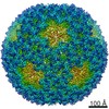 0299MC 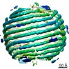 0294C 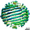 0295C 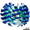 0296C 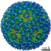 0300C 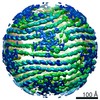 0302C 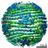 0304C 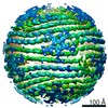 0305C 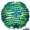 0306C M: map data used to model this data C: citing same article ( |
|---|---|
| Similar structure data |
-Links
 PDBj
PDBj
- Assembly
| Deposited unit | 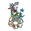
|
|---|---|
| 1 | x 60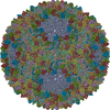
|
| Symmetry | Point symmetry: (Schoenflies symbol: I (icosahedral)) |
-Components
| #1: Protein | Mass: 85080.711 Da / Num. of mol.: 2 / Source method: isolated from a natural source / Source: (natural) Pseudomonas phage phi6 (bacteriophage) / References: UniProt: P11126#2: Protein | | Mass: 35198.426 Da / Num. of mol.: 1 / Source method: isolated from a natural source / Source: (natural) Pseudomonas phage phi6 (bacteriophage)#3: Protein | Mass: 16018.418 Da / Num. of mol.: 10 / Source method: isolated from a natural source / Source: (natural) Pseudomonas phage phi6 (bacteriophage) |
|---|
-Experimental details
-Experiment
| Experiment | Method: ELECTRON MICROSCOPY |
|---|---|
| EM experiment | Aggregation state: PARTICLE / 3D reconstruction method: single particle reconstruction |
- Sample preparation
| Component | Name: Pseudomonas phage phi6Pseudomonas virus phi6 / Type: VIRUS / Entity ID: all / Source: NATURAL | ||||||||||||
|---|---|---|---|---|---|---|---|---|---|---|---|---|---|
| Source (natural) | Organism: Pseudomonas phage phi6 (bacteriophage) | ||||||||||||
| Details of virus | Empty: NO / Enveloped: YES / Isolate: SPECIES / Type: VIRION | ||||||||||||
| Natural host | Organism: Pseudomonas syringae | ||||||||||||
| Virus shell |
| ||||||||||||
| Buffer solution | pH: 7.2 | ||||||||||||
| Specimen | Embedding applied: NO / Shadowing applied: NO / Staining applied: NO / Vitrification applied: YES | ||||||||||||
| Vitrification | Cryogen name: ETHANE |
- Electron microscopy imaging
| Experimental equipment |  Model: Titan Krios / Image courtesy: FEI Company |
|---|---|
| Microscopy | Model: FEI TITAN KRIOS |
| Electron gun | Electron source: FIELD EMISSION GUN / Accelerating voltage: 300 kV / Illumination mode: FLOOD BEAM |
| Electron lens | Mode: BRIGHT FIELDBright-field microscopy |
| Image recording | Electron dose: 33 e/Å2 / Film or detector model: FEI FALCON II (4k x 4k) |
- Processing
| Software | Name: PHENIX / Version: 1.13_2998: / Classification: refinement | ||||||||||||||||||||||||
|---|---|---|---|---|---|---|---|---|---|---|---|---|---|---|---|---|---|---|---|---|---|---|---|---|---|
| EM software |
| ||||||||||||||||||||||||
| CTF correction | Type: PHASE FLIPPING AND AMPLITUDE CORRECTION | ||||||||||||||||||||||||
| Symmetry | Point symmetry: I (icosahedral) | ||||||||||||||||||||||||
| 3D reconstruction | Resolution: 3.5 Å / Resolution method: FSC 0.143 CUT-OFF / Num. of particles: 55265 / Symmetry type: POINT | ||||||||||||||||||||||||
| Atomic model building | Protocol: RIGID BODY FIT | ||||||||||||||||||||||||
| Atomic model building | PDB-ID: 5MUU |