 Movie
Movie Controller

[English] 日本語
 Yorodumi
- PDB-6b00: Thermostabilized mutant of human carbonic anhydrase II - A65T L10... -
+ Open data

- Basic information
| Entry | Database: PDB / ID: 6b00 | ||||||
|---|---|---|---|---|---|---|---|
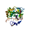
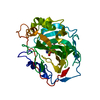
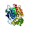
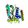
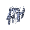
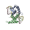
| Title | Thermostabilized mutant of human carbonic anhydrase II - A65T L100H K154N L224S L240P A248T | ||||||
 Components Components | Carbonic anhydrase 2 | ||||||
 Keywords Keywords | LYASE / carbon sequestration / thermostable / protein engineering | ||||||
| Function / homology |  Function and homology information Function and homology informationpositive regulation of cellular pH reduction / positive regulation of dipeptide transmembrane transport / regulation of monoatomic anion transport / secretion / cyanamide hydratase / cyanamide hydratase activity / arylesterase activity / regulation of chloride transport / Reversible hydration of carbon dioxide / angiotensin-activated signaling pathway ...positive regulation of cellular pH reduction / positive regulation of dipeptide transmembrane transport / regulation of monoatomic anion transport / secretion / cyanamide hydratase / cyanamide hydratase activity / arylesterase activity / regulation of chloride transport / Reversible hydration of carbon dioxide / angiotensin-activated signaling pathway / positive regulation of synaptic transmission, GABAergic / morphogenesis of an epithelium / regulation of intracellular pH / carbonic anhydrase / carbonate dehydratase activity / carbon dioxide transport / Erythrocytes take up oxygen and release carbon dioxide / Erythrocytes take up carbon dioxide and release oxygen / neuron cellular homeostasis / one-carbon metabolic process / apical part of cell / myelin sheath / extracellular exosome / zinc ion binding / plasma membrane / cytosol / cytoplasmSimilarity search - Function | ||||||
| Biological species |  Homo sapiens (human) Homo sapiens (human) | ||||||
| Method | X-RAY DIFFRACTION / SYNCHROTRON / MOLECULAR REPLACEMENT / Resolution: 0.9 Å | ||||||
 Authors Authors | Kean, K.M. / Karplus, P.A. | ||||||
 Citation Citation | Journal: Protein Sci. / Year: 2018 Title: Structural insights into a thermostable variant of human carbonic anhydrase II. Authors: Kean, K.M. / Porter, J.J. / Mehl, R.A. / Karplus, P.A. | ||||||
| History |
|
- Structure visualization
| Structure viewer | Molecule: MolmilJmol/JSmol |
|---|
- Downloads & links
-Download
| PDBx/mmCIF format | 6b00.cif.gz | 198.7 KB | Display | PDBx/mmCIF format |
|---|---|---|---|---|
| PDB format | pdb6b00.ent.gz | 160.2 KB | Display | PDB format |
| PDBx/mmJSON format | 6b00.json.gz | Tree view | PDBx/mmJSON format | |
| Others |  Other downloads Other downloads |
-Validation report
| Arichive directory | https://data.pdbj.org/pub/pdb/validation_reports/b0/6b00ftp://data.pdbj.org/pub/pdb/validation_reports/b0/6b00 | HTTPS FTP |
|---|
-Related structure data
| Related structure data |  1ca2S S: Starting model for refinement |
|---|---|
| Similar structure data |
-Links
 PDBj
PDBj

- Assembly
| Deposited unit | 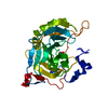
| ||||||||
|---|---|---|---|---|---|---|---|---|---|
| 1 |
| ||||||||
| Unit cell |
|
-Components
| #1: Protein | / Carbonate dehydratase II / Carbonic anhydrase C / CAC / Carbonic anhydrase II / CA-II Mass: 30129.773 Da / Num. of mol.: 1 / Mutation: A65T, L100H, K153N, L223S, L239P, A247T Source method: isolated from a genetically manipulated source Source: (gene. exp.) Homo sapiens (human) / Gene: CA2 / Production host:  Escherichia coli (E. coli) / References: UniProt: P00918, carbonic anhydrase Escherichia coli (E. coli) / References: UniProt: P00918, carbonic anhydrase |
|---|---|
| #2: Chemical | ChemComp-GOL / Glycerol  Mass: 92.094 Da / Num. of mol.: 1 / Source method: obtained synthetically / Formula: C3H8O3 Mass: 92.094 Da / Num. of mol.: 1 / Source method: obtained synthetically / Formula: C3H8O3 |
| #3: Chemical | ChemComp-ZN /   Mass: 65.409 Da / Num. of mol.: 1 / Source method: isolated from a natural source / Formula: Zn Mass: 65.409 Da / Num. of mol.: 1 / Source method: isolated from a natural source / Formula: Zn |
| #4: Water | ChemComp-HOH / Water Mass: 18.015 Da / Num. of mol.: 409 / Source method: isolated from a natural source / Formula: H2O Mass: 18.015 Da / Num. of mol.: 409 / Source method: isolated from a natural source / Formula: H2O |
-Experimental details
-Experiment
| Experiment | Method: X-RAY DIFFRACTION / Number of used crystals: 5 |
|---|
- Sample preparation
| Crystal |
| ||||||||||||||||||||||||
|---|---|---|---|---|---|---|---|---|---|---|---|---|---|---|---|---|---|---|---|---|---|---|---|---|---|
| Crystal grow |
|
-Data collection
| Diffraction |
| ||||||||||||||||||||||||||||||||||||||||||||||||
|---|---|---|---|---|---|---|---|---|---|---|---|---|---|---|---|---|---|---|---|---|---|---|---|---|---|---|---|---|---|---|---|---|---|---|---|---|---|---|---|---|---|---|---|---|---|---|---|---|---|
| Diffraction source |
| ||||||||||||||||||||||||||||||||||||||||||||||||
| Detector |
| ||||||||||||||||||||||||||||||||||||||||||||||||
| Radiation |
| ||||||||||||||||||||||||||||||||||||||||||||||||
| Radiation wavelength |
| ||||||||||||||||||||||||||||||||||||||||||||||||
| Reflection | Entry-ID: 6B00
|

- Processing
| Software |
| |||||||||||||||||||||||||||||||||||||||||||||||||||||||||||||||||||||||||||||||||||||||||||||||||||||||||||||||||||||||
|---|---|---|---|---|---|---|---|---|---|---|---|---|---|---|---|---|---|---|---|---|---|---|---|---|---|---|---|---|---|---|---|---|---|---|---|---|---|---|---|---|---|---|---|---|---|---|---|---|---|---|---|---|---|---|---|---|---|---|---|---|---|---|---|---|---|---|---|---|---|---|---|---|---|---|---|---|---|---|---|---|---|---|---|---|---|---|---|---|---|---|---|---|---|---|---|---|---|---|---|---|---|---|---|---|---|---|---|---|---|---|---|---|---|---|---|---|---|---|---|---|
| Refinement | Method to determine structure: MOLECULAR REPLACEMENT Starting model: 1CA2 Resolution: 0.9→46.788 Å / SU ML: 0.15 / Cross valid method: FREE R-VALUE / σ(F): 1.33 / Phase error: 18.43 / Stereochemistry target values: ML
| |||||||||||||||||||||||||||||||||||||||||||||||||||||||||||||||||||||||||||||||||||||||||||||||||||||||||||||||||||||||
| Solvent computation | Shrinkage radii: 0.9 Å / VDW probe radii: 1.11 Å / Solvent model: FLAT BULK SOLVENT MODEL | |||||||||||||||||||||||||||||||||||||||||||||||||||||||||||||||||||||||||||||||||||||||||||||||||||||||||||||||||||||||
| Refinement step | Cycle: LAST / Resolution: 0.9→46.788 Å
| |||||||||||||||||||||||||||||||||||||||||||||||||||||||||||||||||||||||||||||||||||||||||||||||||||||||||||||||||||||||
| Refine LS restraints |
| |||||||||||||||||||||||||||||||||||||||||||||||||||||||||||||||||||||||||||||||||||||||||||||||||||||||||||||||||||||||
| LS refinement shell |
|
