 Movie
Movie Controller


+ Open data

- Basic information
| Entry | Database: PDB / ID: 5y3r | |||||||||||||||
|---|---|---|---|---|---|---|---|---|---|---|---|---|---|---|---|---|
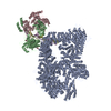
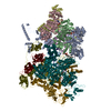
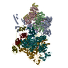
| Title | Cryo-EM structure of Human DNA-PK Holoenzyme | |||||||||||||||
 Components Components |
| |||||||||||||||
 Keywords Keywords | DNA BINDING PROTEIN / Cryo-EM structure / DNA-PK / DNAPKcs / activation / NHEJ | |||||||||||||||
| Function / homology |  Function and homology information Function and homology informationKu70:Ku80 complex / negative regulation of t-circle formation / positive regulation of platelet formation / DNA end binding / T cell receptor V(D)J recombination / pro-B cell differentiation / small-subunit processome assembly / positive regulation of lymphocyte differentiation / DNA-dependent protein kinase activity / histone H2AXS139 kinase activity ...Ku70:Ku80 complex / negative regulation of t-circle formation / positive regulation of platelet formation / DNA end binding / T cell receptor V(D)J recombination / pro-B cell differentiation / small-subunit processome assembly / positive regulation of lymphocyte differentiation / DNA-dependent protein kinase activity / histone H2AXS139 kinase activity / DNA-dependent protein kinase complex / immature B cell differentiation / DNA-dependent protein kinase-DNA ligase 4 complex / cellular response to X-ray / immunoglobulin V(D)J recombination / nonhomologous end joining complex / DNA ligation / regulation of smooth muscle cell proliferation / nuclear telomere cap complex / Cytosolic sensors of pathogen-associated DNA / double-strand break repair via classical nonhomologous end joining / regulation of epithelial cell proliferation / IRF3-mediated induction of type I IFN / telomere capping / recombinational repair / regulation of hematopoietic stem cell differentiation / regulation of telomere maintenance / U3 snoRNA binding / protein localization to chromosome, telomeric region / cellular response to fatty acid / positive regulation of neurogenesis / hematopoietic stem cell proliferation / cellular hyperosmotic salinity response / positive regulation of catalytic activity / T cell lineage commitment / negative regulation of cGAS/STING signaling pathway / telomeric DNA binding / double-strand break repair via alternative nonhomologous end joining / maturation of 5.8S rRNA / B cell lineage commitment / 2-LTR circle formation / positive regulation of double-strand break repair via nonhomologous end joining / mitotic G1 DNA damage checkpoint signaling / : / site of DNA damage / Lyases; Carbon-oxygen lyases; Other carbon-oxygen lyases / cyclin binding / 5'-deoxyribose-5-phosphate lyase activity / hematopoietic stem cell differentiation / positive regulation of protein kinase activity / ectopic germ cell programmed cell death / ATP-dependent activity, acting on DNA / neurogenesis / somitogenesis / enzyme activator activity / positive regulation of telomere maintenance via telomerase / activation of innate immune response / DNA helicase activity / telomere maintenance / positive regulation of erythrocyte differentiation / negative regulation of protein phosphorylation / cellular response to leukemia inhibitory factor / Nonhomologous End-Joining (NHEJ) / small-subunit processome / positive regulation of translation / regulation of circadian rhythm / protein-DNA complex / response to gamma radiation / peptidyl-threonine phosphorylation / Hydrolases; Acting on acid anhydrides; Acting on acid anhydrides to facilitate cellular and subcellular movement / protein destabilization / protein modification process / brain development / cellular response to insulin stimulus / cellular response to gamma radiation / double-strand break repair via nonhomologous end joining / rhythmic process / intrinsic apoptotic signaling pathway in response to DNA damage / double-strand break repair / E3 ubiquitin ligases ubiquitinate target proteins / double-stranded DNA binding / heart development / peptidyl-serine phosphorylation / chromosome, telomeric region / T cell differentiation in thymus / scaffold protein binding / transcription regulator complex / secretory granule lumen / DNA recombination / RNA polymerase II-specific DNA-binding transcription factor binding / ficolin-1-rich granule lumen / transcription cis-regulatory region binding / damaged DNA binding / non-specific serine/threonine protein kinase / protein kinase activity / ribonucleoprotein complex / response to xenobiotic stimulus / protein domain specific binding / positive regulation of apoptotic process / protein phosphorylation Similarity search - Function | |||||||||||||||
| Biological species |  Homo sapiens (human) Homo sapiens (human) | |||||||||||||||
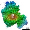
| Method | ELECTRON MICROSCOPY / single particle reconstruction / cryo EM / Resolution: 6.6 Å | |||||||||||||||
 Authors Authors | Yin, X. / Liu, M. / Tian, Y. / Wang, J. / Xu, Y. | |||||||||||||||
| Funding support |  China, 4items China, 4items
| |||||||||||||||
 Citation Citation | Journal: Cell Res / Year: 2017 Title: Cryo-EM structure of human DNA-PK holoenzyme. Authors: Xiaotong Yin / Mengjie Liu / Yuan Tian / Jiawei Wang / Yanhui Xu / Abstract: DNA-dependent protein kinase (DNA-PK) is a serine/threonine protein kinase complex composed of a catalytic subunit (DNA-PKcs) and KU70/80 heterodimer bound to DNA. DNA-PK holoenzyme plays a critical ...DNA-dependent protein kinase (DNA-PK) is a serine/threonine protein kinase complex composed of a catalytic subunit (DNA-PKcs) and KU70/80 heterodimer bound to DNA. DNA-PK holoenzyme plays a critical role in non-homologous end joining (NHEJ), the major DNA repair pathway. Here, we determined cryo-electron microscopy structure of human DNA-PK holoenzyme at 6.6 Å resolution. In the complex structure, DNA-PKcs, KU70, KU80 and DNA duplex form a 650-kDa heterotetramer with 1:1:1:1 stoichiometry. The N-terminal α-solenoid (∼2 800 residues) of DNA-PKcs adopts a double-ring fold and connects the catalytic core domain of DNA-PKcs and KU70/80-DNA. DNA-PKcs and KU70/80 together form a DNA-binding tunnel, which cradles ∼30-bp DNA and prevents sliding inward of DNA-PKcs along with DNA duplex, suggesting a mechanism by which the broken DNA end is protected from unnecessary processing. Structural and biochemical analyses indicate that KU70/80 and DNA coordinately induce conformational changes of DNA-PKcs and allosterically stimulate its kinase activity. We propose a model for activation of DNA-PKcs in which allosteric signals are generated upon DNA-PK holoenzyme formation and transmitted to the kinase domain through N-terminal HEAT repeats and FAT domain of DNA-PKcs. Our studies suggest a mechanism for recognition and protection of broken DNA ends and provide a structural basis for understanding the activation of DNA-PKcs and DNA-PK-mediated NHEJ pathway. | |||||||||||||||
| History |
|
- Structure visualization
| Movie |
Movie viewer |
|---|---|
| Structure viewer | Molecule: MolmilJmol/JSmol |
- Downloads & links
-Download
| PDBx/mmCIF format | 5y3r.cif.gz | 903 KB | Display | PDBx/mmCIF format |
|---|---|---|---|---|
| PDB format | pdb5y3r.ent.gz | 715.1 KB | Display | PDB format |
| PDBx/mmJSON format | 5y3r.json.gz | Tree view | PDBx/mmJSON format | |
| Others |  Other downloads Other downloads |
-Validation report
| Summary document | 5y3r_validation.pdf.gz | 859.5 KB | Display | wwPDB validaton report |
|---|---|---|---|---|
| Full document | 5y3r_full_validation.pdf.gz | 1.4 MB | Display | |
| Data in XML | 5y3r_validation.xml.gz | 211.8 KB | Display | |
| Data in CIF | 5y3r_validation.cif.gz | 308.4 KB | Display | |
| Arichive directory | https://data.pdbj.org/pub/pdb/validation_reports/y3/5y3rftp://data.pdbj.org/pub/pdb/validation_reports/y3/5y3r | HTTPS FTP |
-Related structure data
| Related structure data |  6803MC M: map data used to model this data C: citing same article ( |
|---|---|
| Similar structure data |


-Links
 PDBj
PDBj


- Assembly
| Deposited unit | 
|
|---|---|
| 1 |
|
-Components
-X-ray repair cross-complementing protein ... , 2 types, 2 molecules AB
| #1: Protein | Mass: 57619.352 Da / Num. of mol.: 1 / Fragment: UNP residues 34-534 Source method: isolated from a genetically manipulated source Source: (gene. exp.) Homo sapiens (human) / Gene: XRCC6, G22P1 / Production host: Homo sapiens (human)References: UniProt: P12956, Hydrolases; Acting on acid anhydrides; Acting on acid anhydrides to facilitate cellular and subcellular movement, Lyases; Carbon-oxygen lyases; Other carbon-oxygen lyases |
|---|---|
| #2: Protein | Mass: 60972.969 Da / Num. of mol.: 1 / Fragment: UNP residues 6-541 Source method: isolated from a genetically manipulated source Source: (gene. exp.) Homo sapiens (human) / Gene: XRCC5, G22P2 / Production host: Homo sapiens (human)References: UniProt: P13010, Hydrolases; Acting on acid anhydrides; Acting on acid anhydrides to facilitate cellular and subcellular movement |
-DNA chain , 2 types, 2 molecules DE
| #4: DNA chain | Mass: 10502.785 Da / Num. of mol.: 1 Source method: isolated from a genetically manipulated source Source: (gene. exp.) Homo sapiens (human) / Production host: Homo sapiens (human) |
|---|---|
| #5: DNA chain | Mass: 11042.164 Da / Num. of mol.: 1 Source method: isolated from a genetically manipulated source Source: (gene. exp.) Homo sapiens (human) / Production host: Homo sapiens (human) |
-Protein/peptide / Protein , 2 types, 2 molecules KC
| #3: Protein/peptide | Mass: 1084.182 Da / Num. of mol.: 1 Source method: isolated from a genetically manipulated source Source: (gene. exp.) Homo sapiens (human) / Production host: Homo sapiens (human) |
|---|---|
| #6: Protein | Mass: 468885.344 Da / Num. of mol.: 1 / Fragment: UNP residues 10-4128 Source method: isolated from a genetically manipulated source Source: (gene. exp.) Homo sapiens (human) / Gene: PRKDC, HYRC, HYRC1 / Cell line (production host): HEK293F / Organ (production host): KIDNEY / Production host: Homo sapiens (human)References: UniProt: P78527, non-specific serine/threonine protein kinase |
-Experimental details
-Experiment
| Experiment | Method: ELECTRON MICROSCOPY |
|---|---|
| EM experiment | Aggregation state: PARTICLE / 3D reconstruction method: single particle reconstruction |
- Sample preparation
| Component | Name: PRKDC-Helix / Type: COMPLEX / Entity ID: all / Source: RECOMBINANT |
|---|---|
| Source (natural) | Organism: Homo sapiens (human) |
| Source (recombinant) | Organism: Homo sapiens (human) |
| Buffer solution | pH: 7.4 |
| Specimen | Conc.: 0.6 mg/ml / Embedding applied: NO / Shadowing applied: NO / Staining applied: NO / Vitrification applied: YES |
| Specimen support | Grid material: GOLD / Grid mesh size: 300 divisions/in. / Grid type: Quantifoil R1.2/1.3 |
| Vitrification | Instrument: FEI VITROBOT MARK IV / Cryogen name: ETHANE / Humidity: 100 % / Chamber temperature: 283 K |
- Electron microscopy imaging
| Experimental equipment |  Model: Titan Krios / Image courtesy: FEI Company |
|---|---|
| Microscopy | Model: FEI TITAN KRIOS |
| Electron gun | Electron source:  FIELD EMISSION GUN / Accelerating voltage: 300 kV / Illumination mode: OTHER FIELD EMISSION GUN / Accelerating voltage: 300 kV / Illumination mode: OTHER |
| Electron lens | Mode: BRIGHT FIELD / Nominal magnification: 18000 X / Nominal defocus max: 4700 nm / Nominal defocus min: 1700 nm / Cs: 2.7 mm |
| Specimen holder | Cryogen: NITROGEN / Specimen holder model: FEI TITAN KRIOS AUTOGRID HOLDER |
| Image recording | Average exposure time: 8 sec. / Electron dose: 50 e/Å2 / Detector mode: SUPER-RESOLUTION / Film or detector model: GATAN K2 SUMMIT (4k x 4k) / Num. of real images: 3496 |
| Image scans | Movie frames/image: 32 / Used frames/image: 1-32 |
- Processing
| Software | Name: PHENIX / Version: 1.10.1_2155 / Classification: refinement | ||||||||||||||||||||||||
|---|---|---|---|---|---|---|---|---|---|---|---|---|---|---|---|---|---|---|---|---|---|---|---|---|---|
| EM software |
| ||||||||||||||||||||||||
| CTF correction | Type: NONE | ||||||||||||||||||||||||
| 3D reconstruction | Resolution: 6.6 Å / Resolution method: FSC 0.143 CUT-OFF / Num. of particles: 53451 / Num. of class averages: 1 / Symmetry type: POINT | ||||||||||||||||||||||||
| Refine LS restraints |
|
