 Movie
Movie Controller

[English] 日本語
 Yorodumi
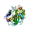
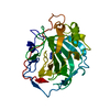
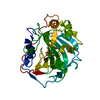
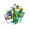
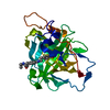
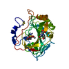
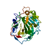
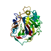
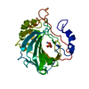
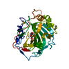
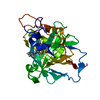
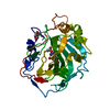
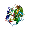
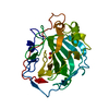
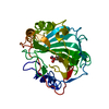
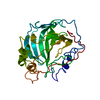
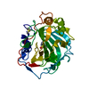
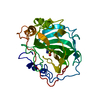
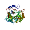
- PDB-5wlr: Carbonic Anhydrase IX-mimic in complex with aryloxy-2-hydroxyprop... -
+ Open data

- Basic information
| Entry | Database: PDB / ID: 5wlr | ||||||
|---|---|---|---|---|---|---|---|
| Title | Carbonic Anhydrase IX-mimic in complex with aryloxy-2-hydroxypropylammine sulfonamide | ||||||
 Components Components | Carbonic Anhydrase IX-mimic | ||||||
 Keywords Keywords | LYASE/INHIBITOR /  carbonic anhydrase / beta adrenergic receptor / sulfonamide / aryloxy-2-hydroxypropylammine / LYASE / LYASE-INHIBITOR complex carbonic anhydrase / beta adrenergic receptor / sulfonamide / aryloxy-2-hydroxypropylammine / LYASE / LYASE-INHIBITOR complex | ||||||
| Function / homology |  Function and homology information Function and homology informationpositive regulation of cellular pH reduction / positive regulation of dipeptide transmembrane transport / regulation of monoatomic anion transport / secretion / cyanamide hydratase / cyanamide hydratase activity / arylesterase activity / regulation of chloride transport / Reversible hydration of carbon dioxide / angiotensin-activated signaling pathway ...positive regulation of cellular pH reduction / positive regulation of dipeptide transmembrane transport / regulation of monoatomic anion transport / secretion / cyanamide hydratase / cyanamide hydratase activity / arylesterase activity / regulation of chloride transport / Reversible hydration of carbon dioxide / angiotensin-activated signaling pathway / positive regulation of synaptic transmission, GABAergic / morphogenesis of an epithelium / regulation of intracellular pH / carbonic anhydrase / carbonate dehydratase activity / carbon dioxide transport / Erythrocytes take up oxygen and release carbon dioxide / Erythrocytes take up carbon dioxide and release oxygen / neuron cellular homeostasis / one-carbon metabolic process / apical part of cell / myelin sheath / extracellular exosome / zinc ion binding / plasma membrane / cytosol / cytoplasmSimilarity search - Function | ||||||
| Biological species |  Homo sapiens (human) Homo sapiens (human) | ||||||
| Method | X-RAY DIFFRACTION / SYNCHROTRON / MOLECULAR REPLACEMENT / Resolution: 1.49 Å | ||||||
 Authors Authors | Lomelino, C.L. / Andring, J.T. / McKenna, R. | ||||||
 Citation Citation | Journal: To Be Published Title: Hybrids for multitargeted therapy Beta Adrenergic Receptor modulators CAIs Authors: Nocentini, A. / Ceruso, M. / Bua, S. / Lomelino, C.L. / Andring, J.T. / McKenna, R. / Lanzi, C. / Masini, E. / Pecori, R. / Matucci, R. / Filippi, L. / Gratteri, P. / Carta, F. / Selleri, S. / Supuran, C.T. | ||||||
| History |
|
- Structure visualization
| Structure viewer | Molecule: MolmilJmol/JSmol |
|---|
- Downloads & links
-Download
| PDBx/mmCIF format | 5wlr.cif.gz | 128.9 KB | Display | PDBx/mmCIF format |
|---|---|---|---|---|
| PDB format | pdb5wlr.ent.gz | 97.3 KB | Display | PDB format |
| PDBx/mmJSON format | 5wlr.json.gz | Tree view | PDBx/mmJSON format | |
| Others |  Other downloads Other downloads |
-Validation report
| Arichive directory | https://data.pdbj.org/pub/pdb/validation_reports/wl/5wlrftp://data.pdbj.org/pub/pdb/validation_reports/wl/5wlr | HTTPS FTP |
|---|
-Related structure data
| Related structure data |  5wluC  3ks3S S: Starting model for refinement C: citing same article ( |
|---|---|
| Similar structure data |

-Links
 PDBj
PDBj

- Assembly
| Deposited unit | 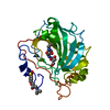
| ||||||||
|---|---|---|---|---|---|---|---|---|---|
| 1 |
| ||||||||
| Unit cell |
|
-Components
| #1: Protein | Mass: 28844.465 Da / Num. of mol.: 1 / Fragment: UNP residues 4-260 Source method: isolated from a genetically manipulated source Source: (gene. exp.) Homo sapiens (human) / Gene: CA2 / Production host:  Escherichia coli (E. coli) / References: UniProt: P00918, carbonic anhydrase Escherichia coli (E. coli) / References: UniProt: P00918, carbonic anhydrase | ||||
|---|---|---|---|---|---|
| #2: Chemical | ChemComp-ZN /   Mass: 65.409 Da / Num. of mol.: 1 / Source method: obtained synthetically / Formula: Zn Mass: 65.409 Da / Num. of mol.: 1 / Source method: obtained synthetically / Formula: Zn | ||||
| #3: Chemical | 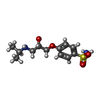  Mass: 288.363 Da / Num. of mol.: 2 / Source method: obtained synthetically / Formula: C12H20N2O4S / Feature type: SUBJECT OF INVESTIGATION Mass: 288.363 Da / Num. of mol.: 2 / Source method: obtained synthetically / Formula: C12H20N2O4S / Feature type: SUBJECT OF INVESTIGATION#4: Chemical | ChemComp-GOL / | Glycerol  Mass: 92.094 Da / Num. of mol.: 1 / Source method: obtained synthetically / Formula: C3H8O3 Mass: 92.094 Da / Num. of mol.: 1 / Source method: obtained synthetically / Formula: C3H8O3#5: Water | ChemComp-HOH / | Water Mass: 18.015 Da / Num. of mol.: 199 / Source method: isolated from a natural source / Formula: H2O Mass: 18.015 Da / Num. of mol.: 199 / Source method: isolated from a natural source / Formula: H2O |
-Experimental details
-Experiment
| Experiment | Method: X-RAY DIFFRACTION / Number of used crystals: 1 |
|---|
- Sample preparation
| Crystal | Density Matthews: 2.16 Å3/Da / Density % sol: 43 % |
|---|---|
| Crystal grow | Temperature: 298 K / Method: vapor diffusion, hanging drop / pH: 7.8 / Details: 1.6M sodium citrate 50mM Tris-HCl |
-Data collection
| Diffraction | Mean temperature: 100 K |
|---|---|
| Diffraction source | Source: SYNCHROTRON / Site: CHESS  / Beamline: F1 / Wavelength: 0.98 Å / Beamline: F1 / Wavelength: 0.98 Å |
| Detector | Type: DECTRIS PILATUS 6M / Detector: PIXEL / Date: Jun 11, 2017 |
| Radiation | Protocol: SINGLE WAVELENGTH / Monochromatic (M) / Laue (L): M / Scattering type: x-ray |
| Radiation wavelength | Wavelength: 0.98 Å / Relative weight: 1 |
| Reflection | Resolution: 1.49→35.2 Å / Num. obs: 60130 / % possible obs: 99.6 % / Redundancy: 4.7 % / Biso Wilson estimate: 13 Å2 / Rpim(I) all: 0.026 / Net I/σ(I): 26.7 |
- Processing
| Software |
| |||||||||||||||||||||||||||||||||||||||||||||||||||||||||||||||||||||||||||||||||||||||||||||||||||||||||||||||||||||||||||||||||||||||||||||||||||
|---|---|---|---|---|---|---|---|---|---|---|---|---|---|---|---|---|---|---|---|---|---|---|---|---|---|---|---|---|---|---|---|---|---|---|---|---|---|---|---|---|---|---|---|---|---|---|---|---|---|---|---|---|---|---|---|---|---|---|---|---|---|---|---|---|---|---|---|---|---|---|---|---|---|---|---|---|---|---|---|---|---|---|---|---|---|---|---|---|---|---|---|---|---|---|---|---|---|---|---|---|---|---|---|---|---|---|---|---|---|---|---|---|---|---|---|---|---|---|---|---|---|---|---|---|---|---|---|---|---|---|---|---|---|---|---|---|---|---|---|---|---|---|---|---|---|---|---|---|
| Refinement | Method to determine structure: MOLECULAR REPLACEMENT Starting model: 3KS3 Resolution: 1.49→35.2 Å / SU ML: 0.12 / Cross valid method: FREE R-VALUE / σ(F): 1.37 / Phase error: 18.28
| |||||||||||||||||||||||||||||||||||||||||||||||||||||||||||||||||||||||||||||||||||||||||||||||||||||||||||||||||||||||||||||||||||||||||||||||||||
| Solvent computation | Shrinkage radii: 0.9 Å / VDW probe radii: 1.11 Å | |||||||||||||||||||||||||||||||||||||||||||||||||||||||||||||||||||||||||||||||||||||||||||||||||||||||||||||||||||||||||||||||||||||||||||||||||||
| Displacement parameters | Biso max: 113.47 Å2 / Biso mean: 19.5808 Å2 / Biso min: 7.6 Å2 | |||||||||||||||||||||||||||||||||||||||||||||||||||||||||||||||||||||||||||||||||||||||||||||||||||||||||||||||||||||||||||||||||||||||||||||||||||
| Refinement step | Cycle: final / Resolution: 1.49→35.2 Å
| |||||||||||||||||||||||||||||||||||||||||||||||||||||||||||||||||||||||||||||||||||||||||||||||||||||||||||||||||||||||||||||||||||||||||||||||||||
| Refine LS restraints |
| |||||||||||||||||||||||||||||||||||||||||||||||||||||||||||||||||||||||||||||||||||||||||||||||||||||||||||||||||||||||||||||||||||||||||||||||||||
| LS refinement shell | Refine-ID: X-RAY DIFFRACTION / Rfactor Rfree error: 0 / Total num. of bins used: 20
| |||||||||||||||||||||||||||||||||||||||||||||||||||||||||||||||||||||||||||||||||||||||||||||||||||||||||||||||||||||||||||||||||||||||||||||||||||
| Refinement TLS params. | Method: refined / Origin x: -9.3923 Å / Origin y: -1.5654 Å / Origin z: 16.1658 Å
| |||||||||||||||||||||||||||||||||||||||||||||||||||||||||||||||||||||||||||||||||||||||||||||||||||||||||||||||||||||||||||||||||||||||||||||||||||
| Refinement TLS group |
|
