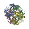
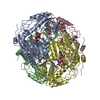
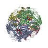
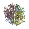
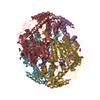
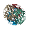
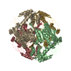
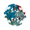
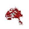
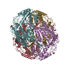
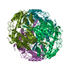
Journal: To be Published Title: Structural and Kinetic Analysis for Cofactor-binding Residues in Mammalian-like Aldehyde Dehydrogenase from Bacillus cereus Involved in Oxidation and Reduction Activity for All-trans-retinal Authors: Ngo, H.P.T. / Hong, S.H. / Oh, D.K. / Kang, L.W.
Resolution: 2.6→19.64 Å / Cor.coef. Fo:Fc: 0.944 / Cor.coef. Fo:Fc free: 0.901 / SU B: 9.187 / SU ML: 0.197 / Cross valid method: THROUGHOUT / ESU R: 1.119 / ESU R Free: 0.294 / Stereochemistry target values: MAXIMUM LIKELIHOOD / Details: HYDROGENS HAVE BEEN USED IF PRESENT IN THE INPUT
Rfactor
Num. reflection
% reflection
Selection details
Rfree
0.22669
3410
5.1 %
RANDOM
Rwork
0.17056
-
-
-
obs
0.17345
63948
99.48 %
-
Solvent computation
Ion probe radii: 0.8 Å / Shrinkage radii: 0.8 Å / VDW probe radii: 1.2 Å / Solvent model: MASK
 Movie
Movie Controller

 Yorodumi
 Open data
 Basic information
 Components
 Keywords
 Function and homology information
 Authors
 Citation
 Structure visualization
 Downloads & links
 Other downloads
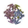


 PDBj
PDBj Assembly
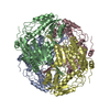

 Mass: 22.990 Da / Num. of mol.: 8 / Source method: obtained synthetically / Formula: Na
 Mass: 18.015 Da / Num. of mol.: 186 / Source method: isolated from a natural source / Formula: H2O
 Sample preparation
 / Beamline: 5C (4A) / Wavelength: 0.97954 Å
 Processing